
Fig. S4
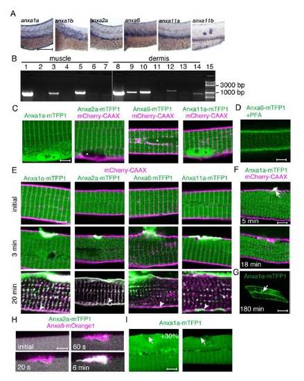
Expression and subcellular localization of annexins in zebrafish. (A) In situ hybridization of annexins anxa1a, anxa1b, anxa2a, anxa6, anxa11a and anxa11b was carried out at 72 hpf (hours post-fertilization). Lateral view of the trunk region is shown. Expression of anxa1a and anxa2a is detectable in the muscle (mRNA enriched at the myoseptal regions) and in the periderm. Anxa6 is highly expressed in the muscle tissue, whereas by 72 hpf only limited amount of anxa11a could be detected in the muscle. Anxa1b and anxa11b expression are limited to the periderm. (B) Reverse transcriptase-PCR was carried out on adult zebrafish skeletal muscle (lanes 1-7) and dermis (lanes 8-14) samples: anxa1a (lanes 1, 8), anxa1b (lanes 2, 9), anxa2a (lanes 3, 10), anxa2b (lanes 4, 11), anxa6 (lanes 5, 12), anxa11a (lanes 6, 13) and anxa11b (7, 14). DNA ladder: 15. Anxa1a, anxa2a and anxa6 are abundant in adult zebrafish muscle tissue. (C) In addition to cytoplasmic localization, strong nuclear signal (asterisk) is visible for Anxa1a and Anxa11a, when fused with mTFP1 (green). Weaker nuclear accumulation is evident for Anxa6-mTFP1 and no nuclear signal could be detected for Anxa2a-mTFP1. mCherry-CAAX marks the cell membrane in magenta. (D) Translocation of Anxa6-mTFP1 to the cell membrane in paraformaldehyde fixed animals. (E) Annexins tagged with mTFP1 (green) accumulate in the sarcolemmal lesion after injury (arrow). Larger cellular wounds eventually lead to the binding of annexins to the membrane distant from the site of injury (arrowhead). (F) Small sarcolemmal wounds lead to temporary accumulation of Anxa1a at the lesion. (G) Large wounds result in persistent Anxa1a localization at the plasma membrane. (H) The relative accumulation kinetics of the studied annexins does not depend on lesion size. Anxa6-mOrange1 (purple) accumulates faster in the large lesion than Anxa2a-mTFP1 (green). Similar results were obtained for small lesions (compare to Figure 4A). (I) Accumulation of annexins does not depend on the expression level. Two cells with 30% difference (largest difference in the study) of Anxa1a-mTFP1 expression were injured. In none of them did the accumulation take place at 60 s after rupture. Scale bars: A, 185 μm; C-F, H-I, 4 μm; G, 65 μm. |
| Genes: | |
|---|---|
| Fish: | |
| Anatomical Terms: | |
| Stage Range: | Protruding-mouth to Adult |
Reprinted from Developmental Cell, 22(3), Roostalu, U., and Strähle, U., In Vivo imaging of molecular interactions at damaged sarcolemma, 515-529, Copyright (2012) with permission from Elsevier. Full text @ Dev. Cell