FIGURE

Fig. 3

ID
ZDB-FIG-120329-62
Publication
Roostalu et al., 2012 - In Vivo imaging of molecular interactions at damaged sarcolemma
Other Figures
All Figure Page
Back to All Figure Page
Fig. 3

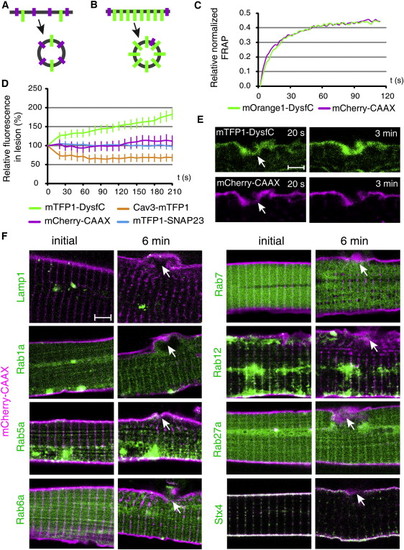
Origin of the Repair Membrane
(A and B) Schematic representation of the two possible sources of the sarcolemmal resealing membrane. (A) Random plasma membrane fragments, decorated with mCherry-CAAX (magenta) or Dysf (green) could provide substrate for membrane repair. (B) Specific Dysf-enriched membrane domains exist for resealing of lesions.
(C) FRAP experiment with mOrange1-tagged DysfC and mCherry-CAAX show comparable kinetics in undamaged myofibers.
(D) The TM domain of Dysf (DysfC) accumulates rapidly at the site of lesion. Much slower or no accumulation was observed for mCherry-CAAX, mTFP1-SNAP23 and Cav3-mTFP1.
(E) Coexpression of mTFP1-DysfC and mCherry-CAAX. In contrast to mTFP1-DysfC (green), mCherry-CAAX (magenta) does not accumulate in the lesion.
(F) Visualization of Lamp1-mTFP1, mTFP1-Rab1a, mTFP1-Rab5a, mTFP1-Rab6a, mTFP1-Rab7, mTFP1-Rab12, mTFP1-Rab27a, and mTFP1-Stx4 localization (green) in uninjured and injured cells. No significant relocation of the studied vesicle markers could be observed. mCherry-CAAX marks the cell membrane in magenta. White arrow indicates damaged sarcolemma.
Scale bars represent 3 μm (E) and 4 μm (F). See also Figures S2 and S3.

Expression Data

Expression Detail
Antibody Labeling
Phenotype Data

Phenotype Detail
Acknowledgments
This image is the copyrighted work of the attributed author or publisher, and ZFIN has permission only to display this image to its users. Additional permissions should be obtained from the applicable author or publisher of the image.

Reprinted from Developmental Cell, 22(3), Roostalu, U., and Strähle, U., In Vivo imaging of molecular interactions at damaged sarcolemma, 515-529, Copyright (2012) with permission from Elsevier. Full text @ Dev. Cell