
Fig. 6
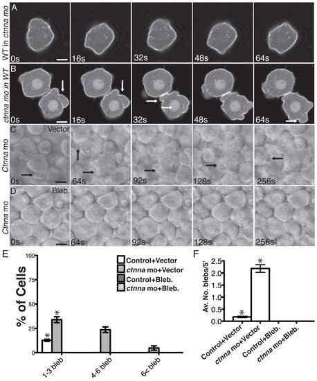
Membrane blebbing triggered by αE-catenin depletion is cell autonomous and myosin II-dependent. (A,B) Selected confocal images from time-lapse movies of transplanted cells expressing mbGFP in the most external epiblast external layer of the host embryos at 50% epiboly. Arrows indicate the blebs. (A) Wild-type (WT) cell in ctnna morphant host embryo. (B) ctnna morphant cell in WT host embryo. Scale bars: 5 μm. (C,D) DIC images from time-lapse movies of untreated (DMSO) embryos or embryos treated with blebbistatin (50 μM) imaged at the most external epiblast layer at the animal pole at 50% epiboly. Scale bars: 8 μm. (E) Percentage of cells exhibiting bleb-like protrusions in untreated embryos or embryos treated with blebbistatin. n=12 embryos. *P<10–8, #P<10–5. Note that no cell exhibited membrane blebbing in embryos treated with blebbistatin. Error bars indicate s.e.m. (F) Average number of blebs per cell untreated embryos or embryos treated with blebbistatin. n=12 embryos. *P<10–9. Note that no cell exhibited membrane blebbing in embryos treated with blebbistatin. Error bars indicate s.e.m. |
| Fish: | |
|---|---|
| Condition: | |
| Knockdown Reagent: | |
| Observed In: | |
| Stage: | 50%-epiboly |