
Fig. S3
- ID
- ZDB-FIG-110812-11
- Publication
- Paridaen et al., 2011 - The nucleolar GTP-binding proteins Gnl2 and nucleostemin are required for retinal neurogenesis in developing zebrafish
- Other Figures
- All Figure Page
- Back to All Figure Page
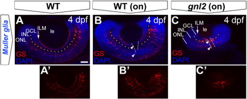
Disorganized Müller glial cells in the gnl2 retina. Single z-plane of whole-mount embryos at 4 dpf labeled with antibody to glutamine synthase, outlining the soma and processes in WT (A and B) versus gnl2 mutant (C). The Müller glial endfeet are ending at the inner limiting membrane (arrow). In gnl2 mutants, the Müller glial somata localization in the INL is disorganized. In addition, glutamine synthase-immunoreactive cells are lining the optic nerve head (arrowheads). Dashed lines are outlining the boundary between GCL and INL and the optic nerve head. GCL, ganglion cell layer; INL, inner nuclear layer; ILM, inner limiting membrane; le, lens; ONL, outer nuclear layer. Scalebar 25 μm. |
Reprinted from Developmental Biology, 355(2), Paridaen, J.T., Janson, E., Utami, K.H., Pereboom, T.C., Essers, P.B., van Rooijen, C., Zivkovic, D., and Macinnes, A.W., The nucleolar GTP-binding proteins Gnl2 and nucleostemin are required for retinal neurogenesis in developing zebrafish, 286-301, Copyright (2011) with permission from Elsevier. Full text @ Dev. Biol.