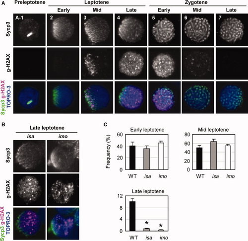
Expression and localization of γ-H2AX in isa and imo mutant spermatocytes. A: Double immunocytochemical analysis of Sycp3 (upper) and γ-H2AX (middle), and counterstaining with TOPRO-3 (blue) in wild-type spermatocytes. No γ-H2AX signals were detected in preleptotene (A-1) and early leptotene (2) cells. (3) At the mid-leptotene stage, γ-H2AX first appeared to be restricted and polarized at the nuclear region in which Sycp3 was clustered. (4) In the late leptotene stage, the γ-H2AX domains are scattered throughout the nucleus as massive accumulations. (5) Early zygotene stage. (6) Mid zygotene stage. (7) Late zygotene stage. γ-H2AX labeling has disappeared. B: Substantial reduction of normal late leptotene spermatocytes in the isa and imo mutants. Mutant leptotene spermatocytes exhibit a severe impairment of γ-H2AX expansion throughout the nucleus. Only a few late leptotene spermatocytes with a scattered expression of γ-H2AX were detectable in isa and imo. C: Quantification of spermatocytes in early, mid, and late leptotene stage spermatocytes. Late leptotene spermatocytes are significantly decreased in mutants. The values shown are the mean ± SEM of 200 leptotene spermatocytes from three zebrafish in each case. Statistical comparisons with wild-type were performed at a given leptotene stage using the Student′s t-test; *P < 0.05.
|

