
Fig. S4
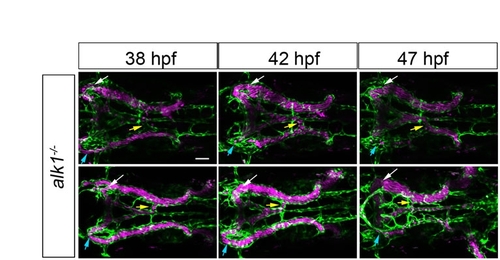
Inter- and intra-embryo variability in AV shunt formation in alk1 mutants. All alk1 mutants initially drain the basal communicating artery via bilateral connections to the primoridal midbrain channel (white and blue arrows), and drain the basilar artery via a variable number of connections to the primordial hindbrain channel (yellow arrows). These connections are also present in wild-type embryos but regress as this arterial system develops (see Fig. 1, main text). By contrast, at least one of these connections is maintained in alk1 mutants. The complement of shunts varies from embryo to embryo, and the prominence of individual shunts in a single embryo changes over time. Each row represents a single alk1-/-; chrna1-/-;Tg(kdrl:GFP)la116;Tg(gata1:dsRed)sd2 embryo imaged at indicated time points. Endothelial cells are green, erythrocytes are magenta. Two-dimensional confocal z-projections, dorsal views, anterior leftwards. Scale bar: 50 μm. |