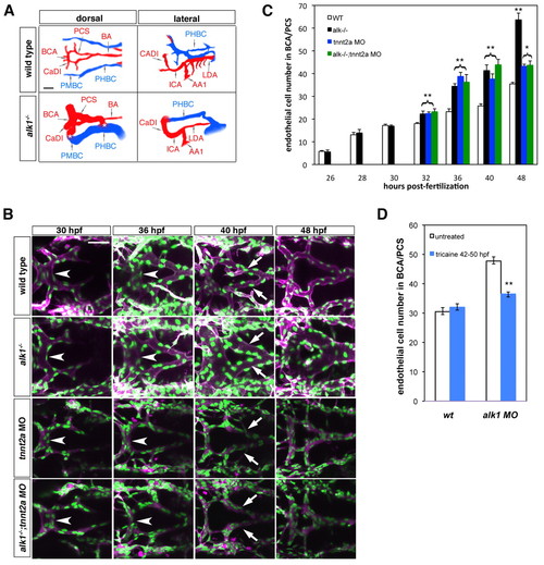
Blood flow modifies endothelial cell number in alk1 mutants. (A) Wiring diagrams of perfused head vessels in 48 hpf wild-type and alk1-/- embryos, derived from two-dimensional confocal projections of Tg(gata1:dsRed)sd2 embryos. Arteries are red; veins are blue. alk1 mutants exhibit enlarged arteries and AVMs (*) between the BCA/PMBC and/or BA/PHBC. Vessels represented in wild-type embryos but not alk1-/- embryos are present but not patent in mutants. AA1, first aortic arch; BA, basilar artery; BCA, basal communicating artery; CaDI, caudal division of internal carotid artery; ICA, internal carotid artery; LDA, lateral dorsal aorta; PCS, posterior communicating segments; PHBC, primordial hindbrain channel; PMBC, primordial midbrain channel. Scale bar: 50 μm. (B) Development of the BCA/PCSs in wild type (row 1), alk1-/- (row 2), tnnt2a morphants (MO) (row 3) and alk1-/-;tnnt2a MO (row 4). Images are two-dimensional confocal projections of live Tg(fli1a:nEGFP)y7;Tg(fli1a.ep:mRFP-F)pt505 embryos, dorsal views, anterior leftwards. Endothelial cell nuclei are green; endothelial cell membranes are magenta. In alk1-/-, the BCA (arrowhead) is enlarged by 36 hpf, and the PCSs (arrows) by 40 hpf. BCA/PCS morphology is relatively normal in tnnt2 MO and alk1-/-;tnnt2 MO, although vessels are collapsed. Scale bar: 50 μm. (C) Quantification of endothelial cell number from confocal micrographs of fixed Tg(fli1a:nEGFP)y7 embryos. alk1-/- with (black bars) or without (green bars) blood flow show similar increases in endothelial cell number compared with wild-type siblings (white bars) between 32-40 hpf, although the pronounced increase between 40 and 48 hpf observed in alk1-/- depends on blood flow. Cell number in tnnt2a MO (blue bars) is not different from cell number in alk1-/-;tnnt2a MO, suggesting that lack of flow phenocopies alk1 mutants in terms of endothelial cell number. Data represent mean±s.e.m. (n=3-13 independent samples) and were analyzed by Student′s t-test. *P<0.01; **P<0.001. (D) Quantification of endothelial cell number from confocal micrographs of 50 hpf wild-type or alk1 MO Tg(fli1a:nEGFP)y7 embryos treated with tricaine between 42 and 50 hpf. The increase in endothelial cell number observed at 40-48 hpf in alk1 morphants is reduced by stopping blood flow. Data represent mean±s.e.m. (n=4-8 independent samples) and were analyzed by Student′s t-test. **P<0.001
|
