
Fig. 3
- ID
- ZDB-FIG-110318-22
- Publication
- Bretaud et al., 2011 - Characterization of spatial and temporal expression pattern of Col15a1b during zebrafish development
- Other Figures
- All Figure Page
- Back to All Figure Page
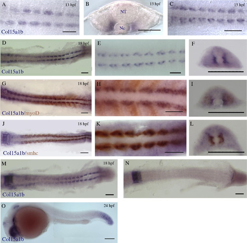
In situ hybridization of Col15a1b antisense RNA probe on whole-mount zebrafish embryos at 13 hpf (8-somite), 15 hpf (12-somite); 18 hpf (18-somite stage), and 24 hpf. (A and C–E) Dorsal views of the trunk region of embryos hybridized with the Col15a1b probe at 13 hpf, 15 hpf and 18 hpf. (B and F) Cross-sections in the trunk region of 8 hpf and 18 hpf-hybridized embryos (Nc: notochord; NT: neural tube). (G and H) Dorsal views and (I) cross-section in the trunk region of an 18 hpf embryo double hybridized with Col15a1b and myoD probes. (J and K) Dorsal views and (L) cross-section in the trunk region of an 18 hpf embryo double hybridized with Col15a1b and smhc probes. (M and N) Dorsal views of 18 hpf embryos hybridized with Col15a1b probe after (N) or not (M) cyclopamine exposure. (O) Lateral view of a 24 hpf embryo hybridized with Col15a1b probe. Scale bar = 100 μm for C, D, F, G, I, J, L, M–O, and 50 μm for A, B, E, H, and K. |
| Gene: | |
|---|---|
| Fish: | |
| Condition: | |
| Anatomical Terms: | |
| Stage Range: | 5-9 somites to Prim-5 |
Reprinted from Gene expression patterns : GEP, 11(1-2), Bretaud, S., Pagnon-Minot, A., Guillon, E., Ruggiero, F., and Le Guellec, D., Characterization of spatial and temporal expression pattern of Col15a1b during zebrafish development, 129-134, Copyright (2011) with permission from Elsevier. Full text @ Gene Expr. Patterns