
Fig. S2
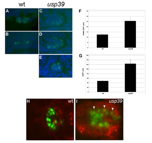
Increase in pituitary cell number in usp39 mutants compared to wt embryos. Immunocytochemistry using anti-GFP antibody was performed on 5 µm frontal sections of wt and usp39 mutants with a POMC-GFP transgenic background at 48 hpf, ventral view, anterior on top. The anti-GFP, rabbit IgG fraction, Alexa Fluor 488 conjugated antibody are at 1:200 dilution (Invitrogen). A drop of ProLong Gold antifade reagent with DAPI (Invitrogen) was added to sections and coverslipped. (A, B) All pituitary POMC-GFP-positive cells are included in two sequential sections of the wild-type embryos. (C–E) Three sections are required to include all pituitary POMC-GFP positive cells in usp39 mutant embryos. (F) Four wt and four usp39 mutants were analyzed for POMC-GFP-positive cells. usp39 mutants had an average of 25.5 POMC-GFP-positive cells compare to 12.5 in wt embryos (mean ± SEM; p<0.02, n = 4). (G) To determine the total number of pituitary cells, DAPI positive cells were counted in the same sections of the four wt and four usp39 mutants. usp39 mutants had an average of 123.5 DAPI-positive cells in the pituitary compare to 48.25 in wt embryos (mean ± SEM; p<0.03, n = 4). (H–I) Embryos were placed in 10 mM solution of BrdU (Sigma) in fish water at 10-somite stage and kept in dark until 48 hpf. The embryos were also injected with 10 nl of 10 mM Brdu at 24 hfp. BrdU labeling results in 48 hpf embryos by immunocytochemistry using anti-GFP antibody and anti-BrdU was performed in whole-mount embryos as described in [Liu et al]. The anti-GFP, rabbit IgG fraction and Alexa Fluor 488 secondary antibody are at 1:200 dilution (Invitrogen). The anti-BrdU, mouse IgG fraction (Santa Cruz) and Alexa Fluor 594 secondary antibody (Invitrogen) are at 1:100 and 1:200 dilution, respectively. Imaging was performed on vibratome 100 μm sections of wt and usp39 mutant embryos. (H) Proliferating cells in the pituitary were not evident in wt embryos. (I) usp39 mutant embryos contain a higher number of proliferating cells in the pituitary region marked by white arrowheads. [Liu NA, Ren M, Song J, Rios Y, Wawrowsky K, et al. (2008) In vivo time-lapse imaging delineates the zebrafish pituitary proopiomelanocortin lineage boundary regulated by FGF3 signal. Dev Biol.] |