|
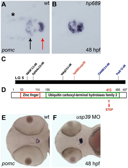
hp689 is a novel zebrafish gene encoding usp39. A, B, E, F: Whole-mount in situ hybridization with pomc probe at 48 hpf, ventral view with anterior to left. (A) Wild-type (wt) embryo, pomc is expressed in the rostral pars distalis (black arrow), the pars intermedia (the red arrow), and in the β-endorphin-synthesizing neurons of the hypothalamus (asterisk). The medial domain which lacks pomc expression corresponds to the proximal pars distalis. (B) The hp689 mutant exhibits increased pomc expression in the adenohypophysis, and lower expression in the hypothalamus compared to wt. (C) Genomic map of linkage group 5 (LG5) and position of the hp689 mutation (in red) and mapping markers based on meiotic segregation linkage analysis. hp689 mapped close to markers z34450 and ndrg3 that were located 2.4 cM and 0.3 cM, respectively (see Materials and Methods). (D) Schematic representation of the Usp39 protein, which include a zinc finger and ubiquitin carboxyl-terminal hydrolases family 2 domains with the hp689 mutation from a tyrosine to a stop codon indicated in red. (E) Non-injected wt control embryos. (F) wt embryos injected with usp39 MO. Note increased pomc expression and disorganization of the adenohypophysis similar to the increased expression of pomc in usp39 mutant embryos in (B).
|