
Fig. 2
- ID
- ZDB-FIG-101115-31
- Publication
- Patten et al., 2009 - PKC{gamma}-induced trafficking of AMPA receptors in embryonic zebrafish depends on NSF and PICK1
- Other Figures
- All Figure Page
- Back to All Figure Page
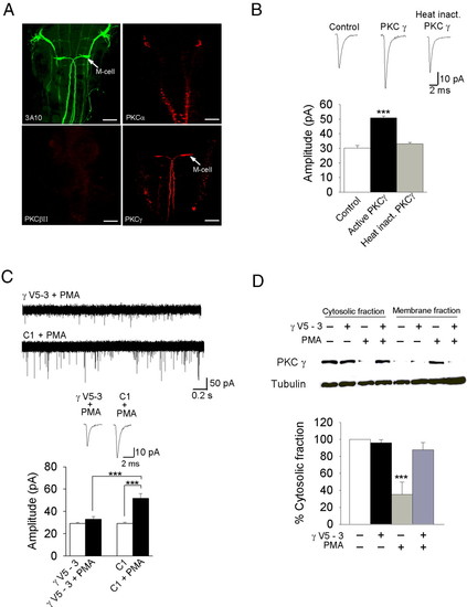
PKCγ is expressed in the Mauthner cell, and its activation leads to an increase in mEPSC amplitude. (A) Anti-3A10, anti-PKCα, anti-βII, and anti-γ immunoreactivity in zebrafish (n = 5). Anti-3A10 is a neurofilament marker that identifies Mauthner cells and is used as a positive control. Only anti-PKCγ labels the Mauthner cell. (Scale bar, 50 μM.) (B) Ten-minutes application of the active form of PKCγ to the Mauthner cell cytosol caused an increase in mEPSC amplitude (n = 5, P < 0.001); 10-min application of heat inactivated PKCγ had no effect (n = 4). Controls represent a recording of AMPA mEPSCs at the 10-min time point in normal intra and extracellular solutions. (C) Intracellular application of γV5-3 (10 nM) blocked the PMA-induced increase in the amplitude (n = 6), whereas the control peptide (C1; 10 nM) had no effect (n = 4). (D) Immunoblot analysis of cytosolic and membrane fractions from zebrafish brain after incubation with or without γV5-3 and PMA (5 nM); γV5-3 inhibited the PMA-induced loss of PKCγ from the cytosolic fraction (n = 4, P < 0.001). ***, significantly different, P < 0.001. |
| Genes: | |
|---|---|
| Antibodies: | |
| Fish: | |
| Anatomical Term: | |
| Stage: | Long-pec |