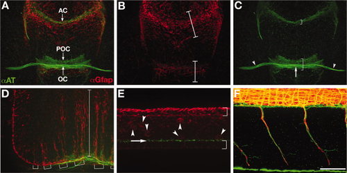
Wild-type axon and astroglial morphology. A-F: Fluorescent immunolabeling of axons (αAT, green) and astroglial cells (αGfap, red) in a 40 hpf wild-type embryo. A-C: Ventral view of the forebrain, anterior up; eyes out of view on sides. A: Combined red and green channels show the association of the forebrain commissures with glial bridges. The anterior commissure forms in the telencephalon and the post optic commissure and optic chiasm form in the diencephalon, which are separated by the optic recess. B: Gfap expressing astroglia are condensed into two “bridges”, one in the telencephalon (upper bracket) and one in the diencephalon (lower bracket; Barresi et al.,2005). C: There are three axonal pathways in the forebrain: the postoptic commissure (lower bracket), anterior commissure (upper bracket), and the optic nerves (arrowheads) that cross the midline to form the optic chiasm (arrow). D: Lateral view of the hindbrain, anterior left, dorsal up. Long radial glial processes are segmentally arranged (brackets) and span from the ventral to dorsal axis of the hindbrain (bracket). E: Lateral view of the spinal cord. This image is focused on the midline/ventricular zone of the spinal cord, with the roofplate to the top and floorplate to the bottom (brackets). Radial glial cell bodies (red, arrowheads) and cilia (green, arrow) are visible just above the floorplate. F: Lateral view of motor nerves in the trunk, anterior left, dorsal up. Axon labeling (green) is distinct from labeling of motor axon associated glia (red). αAT, anti-Aceytlated Tubulin; αGfap, anti-Glial fibrillary acidic protein; tel, (telencephalon; di, diencephalon; POC, postoptic commissure; AC, anterior commissure; OC, optic chiasm. Scale bar = 50 μm.
|

