
Fig. 5
- ID
- ZDB-FIG-101104-1
- Publication
- Lanahan et al., 2010 - VEGF Receptor 2 Endocytic Trafficking Regulates Arterial Morphogenesis
- Other Figures
- All Figure Page
- Back to All Figure Page
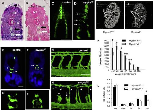
Myo6aKD Impairs Arterial Development in Zebrafish; Functional Effects of Mysosin VI Gene Disruption in Mice (A and B) Transverse section through the trunk of a 28 hpf zebrafish embryo stained by whole-mount in situ hybridization using a myo6a specific antisense (A) or sense (B) riboprobe. Myo6a expression was observed in the neural tube (NT), somites (S), and both the dorsal aorta (black arrow) and posterior cardinal vein (white arrow). The observed expression pattern is specific, as no signal was observed upon hybridization using the sense probe. (C and D) Dorsal view of 16 hpf Tg(fli1:EGFP)y1 embryo, head on top. In control embryos (C), GFP+ angioblasts migrated in an ordered anteroposteriorly directed zipper-like pattern from the lateral plate mesoderm toward the midline, where they assembled into the primitive axial vessels. In contrast, in myo6aKD embryos (D), a portion of angioblasts (arrows) stalled along their lateromedial movement and others failed to maintain their correct stereotyped trajectory, resulting in apparently chaotic migration pattern. (E and F) Transverse sections through the trunk of Tg(fli1:EGFP)y1embryos of 30 hpf, following a whole-mount immunostaining using an anti-GFP antibody (green) and counterstained with DAPI (blue); head on top. Compared to control embryos (E and E′), myo6aKD embryos had a strikingly thinner dorsal aorta (arrow) with an obvious reduced lumen size, while the posterior cardinal vein (arrowhead) remained unaffected (F and F′). (E′) and (F′) are magnifications of the axial vessels in (E) and (F), respectively. (G and H) Lateral view on a trunk segment of Tg(fli1:EGFP)y1embryos at 40 hpf; head to the left. In control embryos (G), ISVs sprouted bilaterally from the dorsal aorta adjacent to the ventral somite boundaries and navigated upwards to the laterodorsal roof of the neural tube, where they split, elongated, and fused to form the DLAV. However, in myo6aKD embryos (H), ISVs often consisted of slender endothelial cells (arrowheads) and/or stalled along their dorsal trajectory (arrows), thereby impairing proper DLAV formation (asteriks). Scale bars represent 10 μm in (A), (B), and (E–F′) and 5 μm in (C), (D), (G), (H). See also Figures S3 and S4. (I and J) Representative reconstructed micro-CT images of whole kidneys (16 μm resolution; n = 3) from age- and gender-matched (I) myosinVI+/+ and (J) myosinVI-/- mice. Note marked reduction in branching in myosinVI-/- mice. (K) Quantitative analysis of micro-CT images indicates a marked decrease in total number of <100 µm diameter vessels in myosinVI-/- mice (white bars) relative to myosinVI+/+ mice (black bars). (Mean ± SEM, *p < 0.05). (L) Quantitative analysis of laser Doppler images indicates significant alterations in hindlimb reperfusion 14 days after femoral artery ligation in myosinVI-/- mice (white bars) relative to myosinVI+/+ mice (black bars). (Mean ± SEM, *p < 0.05). |
| Genes: | |
|---|---|
| Fish: | |
| Knockdown Reagent: | |
| Anatomical Terms: | |
| Stage Range: | 14-19 somites to Prim-25 |
| Fish: | |
|---|---|
| Knockdown Reagent: | |
| Observed In: | |
| Stage Range: | 14-19 somites to Prim-25 |
Reprinted from Developmental Cell, 18(5), Lanahan, A.A., Hermans, K., Claes, F., Kerley-Hamilton, J.S., Zhuang, Z.W., Giordano, F.J., Carmeliet, P., and Simons, M., VEGF Receptor 2 Endocytic Trafficking Regulates Arterial Morphogenesis, 713-724, Copyright (2010) with permission from Elsevier. Full text @ Dev. Cell