|
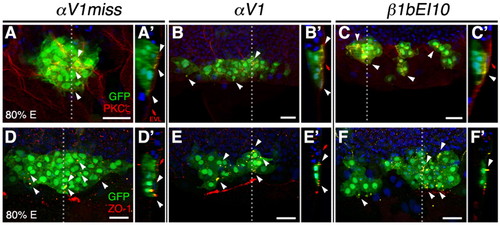
Apical attachment of DFC to EVL is maintained in αV and β1b morphants. (A-F′) Confocal images of DFCs in Tg(sox17:GFP)-expressing embryos (green) nuclear-stained (blue) and immunolabeled with anti-aPKC-ζ antibody (red) (A-C) or with anti-ZO-1 antibody (red) (D-F); channels are merged. Dorsal views of 80% E embryos are shown in all panels, anterior to the top. Sagittal confocal sections at the position of the dotted lines are shown in A′ to F′, embryo surface to the right. Representative embryos that were injected with 1.5 ng αV1miss (A,D), 1.25 ng αV1 (B,E), or 5 ng β1bEI10 (C,F). (A-C) 3D rendering of multiple focal planes through the embryo at the level of DFCs. At 80% E, aPKC-ζ staining demarcates the DFC-EVL interface (A′-C′, white arrowheads). (D-F) Single dorsal focal planes of DFC clusters. Embryos were immunolabeled with ZO-1, which was enriched between DFCs and the interior surface of overlying EVL (D-F′, white arrowheads). Scale bars: 30 μm.
|