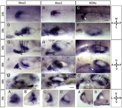
Fig. 1
|
Expression of hmx genes during development of the zebrafish otic vesicle. RNA transcripts were detected using whole mount in situ hybridization. Probes, stages, and orientation are indicated, with the plane of transverse sections (A′–D′ and A″, B″) shown in panels G and I. Both hmx2 (A, D, G, J, M) and hmx3 (B, E, H, K, N) are expressed in the anterior otic vesicle and in the cells anterior to the otic vesicle (arrows). SOHo (C, F, I, L, O) is expressed in the postero-ventral part of the otic vesicle and the anterior lateral line ganglion (white arrowheads). Expression of all three hmx genes is restricted to the area of semicircular canals at later stages. Scale bars: (A–O) 50 μm, (A′–D′, A″, B″) 25 μm. Abbreviations: A, anterior; P, posterior; M, medial; L, lateral; D, dorsal; V, ventral. |
| Genes: | |
|---|---|
| Fish: | |
| Anatomical Terms: | |
| Stage Range: | Prim-5 to Long-pec |
Reprinted from Developmental Biology, 339(2), Feng, Y., and Xu, Q., Pivotal role of hmx2 and hmx3 in zebrafish inner ear and lateral line development, 507-518, Copyright (2010) with permission from Elsevier. Full text @ Dev. Biol.

