
Fig. S5
- ID
- ZDB-FIG-100309-40
- Publication
- Gutzman et al., 2010 - Epithelial relaxation mediated by the myosin phosphatase regulator Mypt1 is required for brain ventricle lumen expansion and hindbrain morphogenesis
- Other Figures
- All Figure Page
- Back to All Figure Page
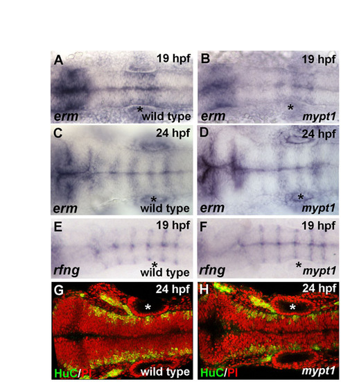
Hindbrain gene expression and neuronal differentiation are normal in mypt1 mutants. Gene expression patterns appeared normal for all marker genes examined in mypt1 mutants versus wild type. (A-D) erm in situ hybridization in wild type (A,C) and mypt1 mutants (B,D) at 19 and 24 hpf, respectively. Gene expression is normal in mypt1 mutants and changes normally over this time period. (E,F) 19 hpf wild-type embryos and mypt1 mutants were analyzed for rfng expression by in situ hybridization. rfng is expressed in rhombomere boundaries in wild-type embryos and was normal in mypt1 mutants. (G,H) Postmitotic neurons were labeled for HuC (green) and nuclei were counterstained with propidium iodide (red). Anterior is to the left in all images. Normal gene expression for wnt1, krox20, pax2a, zic1 and shh was also seen (data not shown). Asterisks indicate ear. |