
Fig. S2
- ID
- ZDB-FIG-100309-37
- Publication
- Gutzman et al., 2010 - Epithelial relaxation mediated by the myosin phosphatase regulator Mypt1 is required for brain ventricle lumen expansion and hindbrain morphogenesis
- Other Figures
- All Figure Page
- Back to All Figure Page
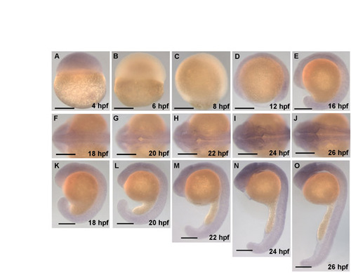
mypt1 is expressed throughout the brain during hindbrain ventricle formation. In situ hybridization showing the expression of mypt1 over time, including 18-24 hpf, during which the hindbrain ventricle forms. The in situ probe was constructed from an mypt1 cDNA fragment from 132 to 3228 bp. Forward primer (from +123 to +144 relative to the start site), 5′-CGAAGGTGAAGTTCGACGATG-3′ and reverse primer (+3228 to +3248), 5′-TGTCGCCATTGTCTCGTGTT-3′. This fragment was subcloned into pGEM using the pGEM T-Easy Vector System Kit (Promega). In situ hybridization was conducted using standard procedures. (A-E) Lateral views at the stages indicated. (F-J) Dorsal views at the stages indicated. (K-O) Lateral views of the corresponding dorsal views from embryos in panels directly above. Scale bars: 200 μm. |