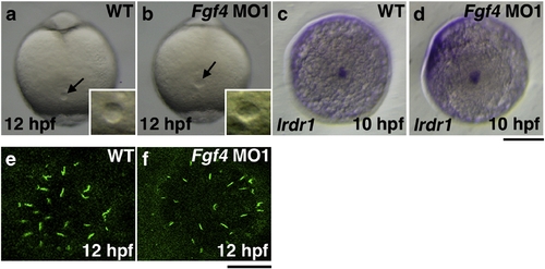
The formation of Kupffer′s vesicle in Fgf4 knockdown embryos. Kupffer′s vesicle in Fgf4 MO1-injected embryos at 12 hpf was morphologically normal (100%, n = 32) (a, b). The expression of lrdr1, which is essential for cilia to function in Kupffer′s vesicle, was examined by whole mount in situ hybridization. The expression was also unaffected in Fgf4 MO1-injected embryos at 10 hpf (93.3%, n = 30) (c, d). (a–d) Dorsal views of the tail bud. A scale bar = 200 μm. Cilia exist on the cells in Kupffer′s vesicle. The expression of acetylated tubulin, a marker protein for cilia, was examined by immunohistochemistry using an anti-acetylated tubulin antibody. The size of Kupffer′s vesicle and the number of cilia of Kupffer′s vesicle in Fgf4 MO-injected embryos were not significantly affected (100%, n = 30); however, the average length of cilia of Kupffer′s vesicle in Fgf4 MO-injected embryos (3.27 ± 1.06 μm, n = 204) was significantly shorter than that in wild-type embryos (4.41. ± 0.91 μm, n = 214) (e, f). These results indicate that Fgf4 is not essential for the morphology and function of Kupffer′s vesicle. A scale bar = 20 μm.
|
