
Fig. 3
- ID
- ZDB-FIG-090324-23
- Publication
- Kimmel et al., 1998 - The shaping of the pharnygeal cartilages during early development of the zebrafish
- Other Figures
- All Figure Page
- Back to All Figure Page
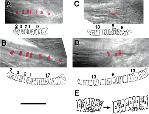
Formation of the SY cell stacks. Two examples (A and B, C–E) are shown. Cells were labeled in the early embryo, by injection with rhodamine–dextran (see Methods and Materials) and the SY, when containing labeled descendants of the injected cells, was photographed in the same two live larvae at 3 days (A, C) and 4 days (B, D) of development. The micrographs show overlays of single-plane images of epifluorescence (with fluorescent labeling pseudo-colored red) and with Nomarski bright-field illumination through the same SY cartilage at the two stages. Anterior is to the left. The accompanying drawings show the SY cell stack reconstructed from the multiple focal plane Nomarski images, with the labeled chondrocytes stippled. In A and B the three labeled cells to the right are perichondrial cells, not chondrocytes, and they are not included in the drawings. The numbers show how many unlabeled cells are distributed along the cartilage relative to the positions of the labeled ones. The dotted cell outlines to the right show a few of the unstacked cells present in the IJ. Between the two time points unlabeled cells add preferentially to the posterior SY (to the right) in these examples, and in another case analyzed similarly. (C) An enlargement of the local region including the labeled cells in the second larva. The number within each cell indicates the number of chondrocyte neighbors it contacts. Less contacts are made at the older stage, presumably due to cellular intercalations occurring between the two times, and that make the more orderly stack at the later time. Scale bar: 50 μm for A–D, 25 μm for E. |
Reprinted from Developmental Biology, 203, Kimmel, C.B., Miller, C.T., Kruse, G., Ullmann, B., BreMiller, R.A., Larison, K.D., and Snyder, H.C., The shaping of the pharnygeal cartilages during early development of the zebrafish, 245-263, Copyright (1998) with permission from Elsevier. Full text @ Dev. Biol.