
Fig. 1
- ID
- ZDB-FIG-090324-21
- Publication
- Kimmel et al., 1998 - The shaping of the pharnygeal cartilages during early development of the zebrafish
- Other Figures
- All Figure Page
- Back to All Figure Page
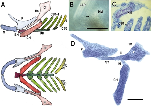
Pharyngeal cartilages and their arrangements of chondrocytes. (A) Diagram of the segmental series of cartilages of the young larva, in a left-side view above (dorsal to the top and anterior to the left) and in a ventral view below. P and M are dorsal and ventral cartilages of the mandibular (first pharyngeal) arch (all abbreviations used in this paper are listed at the end of this legend). HS and CH are their putative homologues in the hyoid (second pharyngeal) arch. CB1-4 bear the set of four gills and CB5 (seventh pharyngeal arch) bears teeth. BH and BB are in the ventral midline (see Schilling and Kimmel, 1997 for a description of all of these cartilages and features of their development). (B) Dorsolateral surface view of the HM. Nomarski imaging in the live 5-day larva, revealing the mosaic-tile appearance of its cells. The arrow points to a nerve that passes through the hole in the HM, prominent in other figures (e.g., D), the hyomandibular foramen. Striated muscle fibers of LAP are also evident. Through-focus sectioning reveals that no chondrocytes are present above or below the plane of focus in the figure; there is only a single layer of them. (C) Horizontal Epon 7.5-μm section through the pharynx at 4 days, stained with methylene blue and basic fuchsin. Cartilage matrix is colored purple, the final magnification is the same as for B. The section passes through the HM, and through CB1–3. The HM cells in such sections have a low-columnar appearance; a view across such a plate of cartilage is rather like a view along a thinner rod of cartilage such as the SY (i.e., the view of the HM here is similar to the view of the SY in D). Sections across the rod-like cartilages, here illustrated for CB1–3, show disk-shaped single cellular profiles, revealing that the cartilages have the structure of a stack of coins or checkers. Irrespective of the form of the cartilages, or how the chondrocytes are arranged, the overlying, immediately adjacent layer of perichondrial cells is flattened onto the cartilage and forms a rather complete sheath. Perichondrial cell nuclei are more elongated and compact than chondrocyte nuclei. (D) Alcian green-stained cartilages, dissected out from a 5-day larva and flat-mounted (see Materials and Methods). The presentation shows all of the chondrocytes present in these cartilages, and their organization. They lie in rows, roughly parallel to the long axes of the cartilages (i.e., the rows run roughly horizontally in P, SY, and HM, and vertically in CH). Rows are not evident in the IJ. The method does not distort the cartilage shapes, but may disturb the articulations and the positional relationships between adjacent cartilages. Hence the in vivo positional relationships of these elements is better shown in A than D. Scale bars: 100 μm. Abbreviations: BB, basibranchial; BH, basihyal; CB1–5, ceratobranchials 1–5; CH, ceratohyal; HM, hyomandibular region of the HS; HS, hyosymplectic; IH, interhyal; IJ, interhyal joint region of the HS; LAP, levator arcus palatini muscle; M, Meckel’s; P, palatoquadrate; SY, symplectic region of the HS. |
Reprinted from Developmental Biology, 203, Kimmel, C.B., Miller, C.T., Kruse, G., Ullmann, B., BreMiller, R.A., Larison, K.D., and Snyder, H.C., The shaping of the pharnygeal cartilages during early development of the zebrafish, 245-263, Copyright (1998) with permission from Elsevier. Full text @ Dev. Biol.