FIGURE

Fig. 1

ID
ZDB-FIG-090304-56
Publication
Feijoo et al., 2009 - Formation of posterior cranial placode derivatives requires the Iroquois transcription factor irx4a
Other Figures
All Figure Page
Back to All Figure Page
Fig. 1

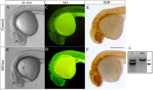
irx4a morphant embryos show defects in head size and extensive cell death. (A, B) Images of live wild type (A) and morphant (B) embryos at 24 hpf; Compare the size of the eyes and head (arrows). (C, D) Acridine orange staining of wild type (C) and irx4a morphant (D) embryos viewed at 24 hpf. Note the increased number of labeled cells in the morphant embryo (E, F) MO4as injected embryos do not show substantial changes in cell proliferation as evidenced by anti-phospho-histone H3 (H3-P) immunostaining. (G) RT-PCR showing the effect of the MO4as on irx4a transcript splicing. RNA prepared from uninjected embryos (“C”) shows the internal control band (actin, act) but the absence of unprocessed irx4a mRNA. The presence of this band in MO4as injected fish (arrow) indicates defective irx4a mRNA splicing. Panels A–F show lateral views of 24 hpf embryos; dorsal is up, anterior is left. Scale bar in F: 70 μm.

Expression Data
Gene:
Antibody:
Fish:
Knockdown Reagent:
Anatomical Term:
Stage: Prim-5

Expression Detail
Antibody Labeling
Phenotype Data
Fish:
Knockdown Reagent:
Observed In:
Stage: Prim-5

Phenotype Detail
Acknowledgments
This image is the copyrighted work of the attributed author or publisher, and ZFIN has permission only to display this image to its users. Additional permissions should be obtained from the applicable author or publisher of the image.

Reprinted from Molecular and cellular neurosciences, 40(3), Feijoo, C.G., Saldias, M.P., De la Paz, J.F., Gómez-Skarmeta, J.L., and Allende, M.L., Formation of posterior cranial placode derivatives requires the Iroquois transcription factor irx4a, 328-337, Copyright (2009) with permission from Elsevier. Full text @ Mol. Cell Neurosci.