
Fig. 6
- ID
- ZDB-FIG-081105-20
- Publication
- Lewis et al., 1999 - Control of muscle cell-type specification in the zebrafish embryo by hedgehog signalling
- Other Figures
- All Figure Page
- Back to All Figure Page
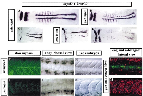
Overexpression of ptc1 suppresses slow muscle specification and MP induction. (A–E) Embryos at early somitogenesis stages and (F–M) embryos at 24 hpf. (B, D, and E) Dorsal flat preparations illustrating the variable effects on adaxial myoD expression following overexpression of ptc1. These range from near elimination of adaxial MyoD expression (E) to a reduction in transcript levels (B) reminiscent of the effects of syu, con, or you mutations (compare with Figs. 2C, 2E, and 2G). The unilateral effects in B and D are most likely due to the unequal distribution of the injected mRNA. (F and I) Lateral view of an uninjected embryo and a sibling embryo injected with ptc1 mRNA stained with an antibody (BA-D5) that recognises slow muscle myosin. Note the strong staining of the muscle fibres and distinct chevron shape of the somites in the uninjected embryo (F). Very few slow muscle fibres are detectable with the BA-D5 antibody in the ptc1-injected embryo at 24 h. Note also the absence of the horizontal myoseptum and the u-shaped somites of this embryo. (G and J) Dorsal views of an uninjected embryo and a sibling embryo injected with ptc1 mRNA, stained with the 4D9 mAb to reveal Eng expression. Note the complete elimination of MPs on one side of the notochord in the ptc1-injected embryo. (H and K) Somites of live embryos at 24 hpf. The embryo injected with ptc1 (K) has u-shaped somites, reminiscent of the u-type mutants; compare with the chevron-shaped somites of the wild-type embryo (H). (L and M) Eng expression in the MPs (green staining) in embryos co-injected with ptc1 and n-βgal mRNAs. In (L) the majority of the injected message (as revealed by distribution of the N-βgal protein, red staining) is localised to the neural tube and there is no effect on MP induction as revealed by wild-type pattern of Eng protein expression in the somites. Also note that the somites are chevron shaped as in wild-type embryos. The low level Eng expression in a cloud of nuclei around the MPs is in the adjoining fast muscle fibres. In the embryo shown in (M), in contrast, there is a large amount of injected message that is localised in the somites as revealed by the distribution of the N-βgal protein. In this case, there is no detectable Eng expression, indicating that MP induction has been inhibited in the Ptc1-expressing somitic cells. Note the u-shaped nature of the somites. |
Reprinted from Developmental Biology, 216(2), Lewis, K.E., Currie, P.D., Roy, S., Schauerte, H., Haffter, P., and Ingham, P.W., Control of muscle cell-type specification in the zebrafish embryo by hedgehog signalling, 469-480, Copyright (1999) with permission from Elsevier. Full text @ Dev. Biol.