
FIGURE
Fig. S1
- ID
- ZDB-FIG-080909-33
- Publication
- Bill et al., 2008 - Development and notch signaling requirements of the zebrafish choroid plexus
- Other Figures
- All Figure Page
- Back to All Figure Page
Fig. S1
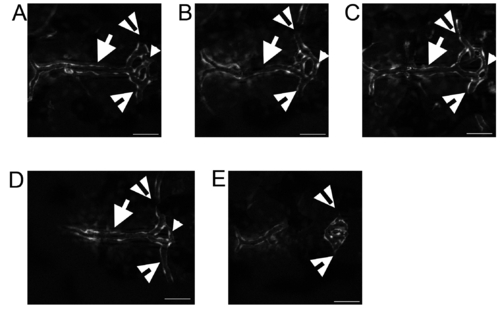
Variation of the DLV-PCeV junction. The DLV (arrows) bifurcated to join the PCeV and PCeV′ (open arrowheads) and the TCB (small arrowheads) connects the two junctions. This structure is plastic. The most common structure includes a third branch that transits between the DLV and the TCB (A–C), but in some instances the third DLV-TCB branch does not form by 5 dpf (D). The most rare occurrence is that the DLV does not develop (E). The PCeV and PCeV′ still meet in the location of the pCP, but lack blood flow (E, open arrowheads). All images are shown with anterior to the left. Scale bar is 50 μm. |
Expression Data
| Gene: | |
|---|---|
| Fish: | |
| Anatomical Terms: | |
| Stage: | Day 5 |
Expression Detail
Antibody Labeling
Phenotype Data
Phenotype Detail
Acknowledgments
This image is the copyrighted work of the attributed author or publisher, and
ZFIN has permission only to display this image to its users.
Additional permissions should be obtained from the applicable author or publisher of the image.
Full text @ PLoS One