
Fig. 8
- ID
- ZDB-FIG-080604-44
- Publication
- Chi et al., 2008 - Genetic and Physiologic Dissection of the Vertebrate Cardiac Conduction System
- Other Figures
- All Figure Page
- Back to All Figure Page
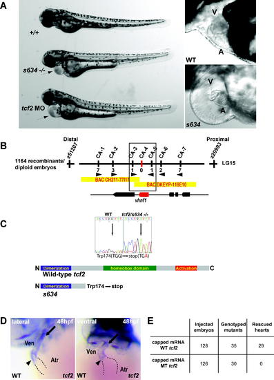
s634 Affects tcf2 (B) Genetic map of the s634 region. Numbers below simple sequence length polymorphism markers indicate recombination events. Two genes were identified within the critical region, which spans two bacterial artificial chromosomes. (C) Sequencing of tcf2s634 cDNA revealed a G to A change at base pair 522, resulting in a premature stop codon at amino acid 174 thereby removing the homeobox and transactivation domains. (D) Whole mount RNA in situ hybridization at 48 hpf reveals tcf2 expression at the AV canal (arrowhead) and the OFT (arrow) region of the ventricle. (E) Injection of wild-type tcf2 mRNA rescued the heart phenotype of ∼82% of s634 mutants, while injection of mutant tcf2 mRNA failed to rescue. A, atrium; V, ventricle. |
| Gene: | |
|---|---|
| Fish: | |
| Anatomical Terms: | |
| Stage: | Long-pec |
| Fish: | |
|---|---|
| Observed In: | |
| Stage: | Long-pec |