
Fig. 6
- ID
- ZDB-FIG-070914-7
- Publication
- Nica et al., 2006 - Eya1 is required for lineage-specific differentiation, but not for cell survival in the zebrafish adenohypophysis
- Other Figures
- All Figure Page
- Back to All Figure Page
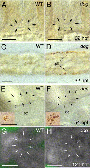
The adenohypophysis of eya1 mutants shows normal apoptosis rates. Panels A-F show TUNEL stainings, panels G, H acridine orange stainings of eya1 mutants and wild-type siblings at stages indicated in lower right corners (hpf; hours post-fertilization). (A, B) Frontal views on heads, dorsal up. (C, D) Dorsal views on tails of same embryos shown in panels A, B, showing apoptosis of cells in the posterior lateral line primordium (pll). (E, F) Lateral views on heads at level of oral cavity (oc). (G, H) Ventral views on heads. Adenohypophysis in panels A, B, E-H is outlined by arrows. Insets in panels E, F show, as internal control, apoptotic cells from inner ear region of same specimen (with more apoptotic cells in mutant; compare with Kozlowski et al., 2005). Scale bars are 50 μm. |
Reprinted from Developmental Biology, 292(1), Nica, G., Herzog, W., Sonntag, C., Nowak, M., Schwarz, H., Zapata, A.G., Hammerschmidt, M., Eya1 is required for lineage-specific differentiation, but not for cell survival in the zebrafish adenohypophysis, 189-204, Copyright (2006) with permission from Elsevier. Full text @ Dev. Biol.