
Fig. 7
- ID
- ZDB-FIG-060606-2
- Publication
- Nica et al., 2006 - Eya1 is required for lineage-specific differentiation, but not for cell survival in the zebrafish adenohypophysis
- Other Figures
- All Figure Page
- Back to All Figure Page
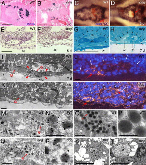
The adenohypophysis of eya1 mutants is of moderately reduced size, with the majority of cells lacking morphological features characteristic for secretion processes. (A, B) Sagittal section after in situ hybridization for pomc, counterstained with eosin/hematoxylin; 3 days post-fertilization (d), anterior to the left. Borders of the pituitary are outlined with dots. (C, D) Double in situ hybridization for prolactin in red and collagen alpha 1(X) in blue, 5 days, ventral view, anterior to the left. prl-positive cells in panel D are indicated with red arrow. (E, F) Longitudinal section stained with eosin/hematoxylene, 5 days, anterior to the left, dorsal to the top. (G, H) Longitudinal section stained with toluidine blue, 7 days, anterior to the left, dorsal to the top. The borders of the pituitary, now with a distinct adenohypophysis (ah) and a neurohypophysis (nh), are indicated by arrows. Note the severe reduction of tissue separating the pituitary from the oral cavity in the mutant (H). (I, K, M–R) Anti-prolactin immunogold electron micrographs of longitudinal sections, anterior to the left, dorsal to the top, 7 days. (I, K) Overviews over entire pituitaries. Red arrowheads in panel I demarcate region with eight type 2 cells in the wild-type pituitary, which are absent in the mutant (K). Panels J, L show adjacent sections of the same specimen as in panels I, K after fluorescent detection of Prl and DAPI staining of nuclei; dorsal borders of the pituitaries are indicated by white arrows. Red arrows in panels J, L point to rostral region with six Prl-positive cells in wild-type (J), three in mutant (L); red arrowhead in panel L indicates region with Prl-positive cells in more lateral sections of the same mutant pituitary, consistent with the randomized and ectopic positions of prl-positive cells in mutant pituitaries revealed via in situ hybridization (compare with Fig. 3H). Panels M, O, Q show higher magnifications of regions indicated in panel I or K, with type 1 cells in panels M, Q, and a type 2 cell in panel O; panels N, P, R show higher magnifications of regions indicated in panels M, O, or Q, revealing electron-dense gold particles in panels N and R, but not in panel P. (S, T) Electron micrographs with ultrastructure of wild-type (S) or mutant (T) adenohypophyseal cell lacking secretory granules. Both cells contain vacuole-like structures, while only the wild-type cell has an elaborated endoplasmatic reticulum. Abbreviations: ad, adenohypophysis; nh, neurohypophysis; n, nucleus; oc, oral cavity. Scale bars are 50 μm for panels A–H, 5 μm for panels I–J, S, T, 0.5 μm for panels M, O, Q, and 0.25 μm for panels N, P, Q. |
| Genes: | |
|---|---|
| Fish: | |
| Anatomical Terms: | |
| Stage Range: | Protruding-mouth to Days 7-13 |
Reprinted from Developmental Biology, 292(1), Nica, G., Herzog, W., Sonntag, C., Nowak, M., Schwarz, H., Zapata, A.G., Hammerschmidt, M., Eya1 is required for lineage-specific differentiation, but not for cell survival in the zebrafish adenohypophysis, 189-204, Copyright (2006) with permission from Elsevier. Full text @ Dev. Biol.