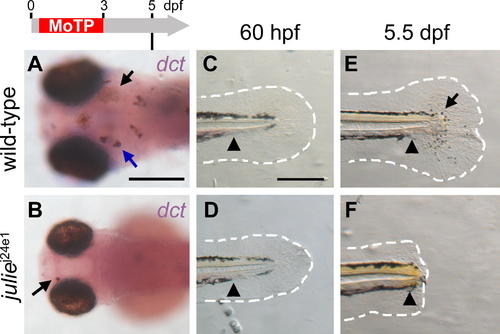
juliej24e1 Mutation Disrupts Melanocyte Regeneration prior to the dct+ Stage and Blocks Tail Regeneration Following Amputation (A and B) Larval melanocytes in juliej24e1 and wild-type larvae were ablated by MoTP treatment from 14 to 72 hpf, and melanocyte regeneration was analyzed at 5 dpf by ISH with dct riboprobes for late-stage melanoblasts. At this stage, many dct+ melanoblasts (melanin-, blue arrow in [A]) and melanocytes (melanin+, black arrow in [A]) appear in wild-type larvae (A). While few melanocytes (melanin+, black arrow in [B]) appear in the juliej24e1 larvae, no dct+ melanoblast was detected by ISH (B). (C–F) juliej24e1 mutants fail to regenerate larval tails following amputation. Larval tails were amputated at 60 hpf (C and D) and tail regeneration in wild-type and juliej24e1 larvae were examined at 5.5 dpf (E and F). At this stage, wild-type larval tails (E) were mostly reconstituted, including fin-fold tissues (outlined with white-dashed line) and melanocytes (arrow in [E]). In the juliej24e1 larvae, the amputation wound healed but the juliej24e1 mutant tail failed to regenerate (F). Arrowheads indicate the amputation planes in (C–F). The white-dashed lines outline the fin folds. Timeline (grey) above (A) indicates the period of MoTP treatment (red) and analysis time (vertical line below the timeline). Scale bars: 250 μm.
|
