
Fig. 4
- ID
- ZDB-FIG-070822-31
- Publication
- Yang et al., 2007 - Mutations in gfpt1 and skiv2l2 Cause Distinct Stage-Specific Defects in Larval Melanocyte Regeneration in Zebrafish
- Other Figures
- All Figure Page
- Back to All Figure Page
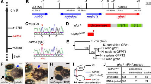
The earthaj23e1 Mutation Is a Nonsense Mutation in gfpt1 (A–E) The earthaj23e1 mutation was mapped to Chromosome 8 between z15786 and z51584 (A). The physical map of the eartha locus was built from the zebrafish genome assembly version 5 (Zv5) with the support of recombination observed in 1,514 meioses. Shown here is a portion of this physical map delimited by seven recombinants at the proximal end and three recombinants at the distal end, containing approximately 189 kb comprising four genes: ntrk2, agtpbp1, mak10, and gfpt1 (B). Sequencing of the earthaj23e1 cDNA for gfpt1 revealed a nonsense mutation in the third exon, where a CAA to TAA substitution resulting in Gln to stop cordon change (C) and (D). The phylogenetic tree of gfpt genes was constructed by using the neighbor-joining method with E. coli glmS as the out-group (E). (F–J) gfpt1 mRNAs partially rescue the eartha phenotypes. gfpt1 mRNAs were in vitro synthesized and injected into one to four cell stage embryos. At 5–7 dpf, 22% of the earthaj23e1 larvae injected with gfpt1 mRNA (J) have some distinctly darker melanocytes (blue arrows in [G]) and more extended pharyngeal skeletons (black arrow in [H] and [I]) than those in the noninjected earthaj23e1 larvae (black and blue arrows in [F]). SIS: sugar isomerase domain. Scale bars: 100 μm. |