|
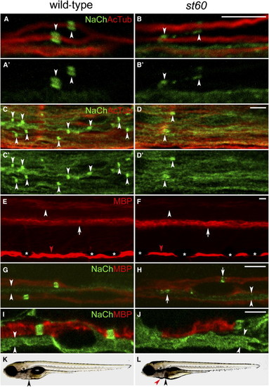
The Nodes of Ranvier Are Abnormal in st60 Mutants (A–B) PLLn of the wild-type (A) and st60 (B) 5 dpf larvae labeled with anti-panNavCh (NaCh) and anti-acetylated tubulin (AcTub). Sodium-channel clusters (indicated by arrowheads) have a distinct morphology in the wild-type (A and A2) but are abnormal in st60 mutants (B and B2). (C–D) Spinal cords of 5 dpf larvae. Sodium-channel clusters (indicated by arrowheads) are more numerous and more regularly shaped in the wild-type (C and C2) than in st60 mutants (D and D2). (E and F) Lateral view of 5 dpf larvae labeled with anti-MBP. Wild-type larvae (E) have myelinated axons in a dorsal region (indicated by arrowhead) and a ventral region (indicated by arrow) of the spinal cord. st60 mutants (F) express a reduced amount of MBP in the dorsal region (indicated by arrowhead), but the ventral region is normal (indicated by arrow). MBP expression in the PLLn is similar in the wild-type and mutant ([E] and [F], indicated by red arrowheads). Visualization of MBP in the PLLn is periodically interrupted by pigment-producing melanophores in both the wild-type and mutants ([E] and [F], indicated by asterisks). (G and H) PLLn of 5 dpf larvae labeled with anti-panNavCh and anti-MBP. Sodium-channel clusters form in the myelinated axons of both the wild-type and mutants, but they are abnormally shaped in st60 mutants ([H], indicated by arrows). Unmyelinated axons have diffuse sodium-channel expression in both the wild-type and mutants ([G] and [H], indicated by arrowheads). (I and J) Motor nerves in 5 dpf larvae labeled with anti-panNavCh and anti-MBP. Mutant motor nerves usually lack MBP, but some have a short region of MBP expression (J) that lacks sodium-channel immunoreactivity. Unmyelinated axons have diffuse sodium-channel expression ([I] and [J], indicated by arrowheads). (K and L) Imaging of live 5 dpf larvae shows that st60 mutants have necrosis in the liver ([K] and [L], indicated by black arrowheads), mild pericardial edema ([L], indicated by red arrowhead), and a failure to inflate the swimbladder. All scale bars represent 5 μm.
|