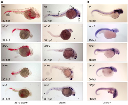
Analysis of zebrafish embryos after MO injections. The developmental stage of each embryo is in the bottom left corner; the targeted gene is indicated in the top left corner. All pictures were taken at the same magnification. (A) Noninjected wild-type (wt) control embryos stained for βE1- (zE1b-globin, left column) and runx1-mRNAs (right column). The ICM, dorsal aorta (DA), primitive erythrocytes (Pr.Ery.), anterior paraxial mesoderm (APM) and olfactory epithelium (OE) are indicated. The βE1signal is red, runx1 signal is blue. Treated embryos were injected with 1 pmol eto-2-MO (32 hpf), 1 pmol cdk9-MO (28 hpf), 1 pmol lmo4-MO (30 hpf), or with 0.5 pmol of the control tcf4-MO (30 hpf). (B) The reduction of runx1 expression in the dorsal aorta region resulting from injection with the MOs directed against eto-2 (n=23/27), cdk9 (n=33/56) and lmo4 (n=36/54) was still observable after 2 days. Embryos injected with the mtgr1-MO were analysed at 38 hpf and showed only a slight effect on the definitive haematopoietic system. All embryos shown were injected with 1 pmol of the corresponding MO.
|
