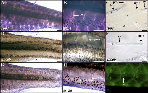
Epidermal expression of rbp4, apoeb, and esr2a in adult zebrafish. A-C: rbp4. D-F: apoeb. G-I: esr2a. A-I: Whole-mount in situ hybridizations with digoxigenin-labeled antisense riboprobes (A,B,D,E,G,H), histological sections after in situ hybridizations (C,F), or in vivo labeling with the fluorescent vital dye 4-di-2-Asp (I) are shown in lateral views and anterior to the left. C,F: Spaces observed between epidermis, scale, and dermis are sectioning artifacts. A,B: Uniform rbp4 hybridization signal distribution is observed in the epidermis of overlapping scales. C: rbp4 is expressed in the basal layer of the epidermis with no signal in epidermal cells covering the anterior and posterior margin regions of the formed scale. D-H: apoeb (D-F) and esr2a (G,H) transcripts are colocalized in the posterior margin epidermis of the scale. G,H: A moderate esr2a hybridization signal is observed in the epidermis covering the whole animal. H: Secondary neuromasts of the lateral lines identified by labeling their hair cells using 4-di-2-Asp (I) express esr2a. ame, anterior margin epidermis of the scale; ce, central epidermis of the scale; d, dermis; n, neuromast; pme, posterior margin epidermis of the scale; s, scale. Scale bars = 500 μm in A,B,D,E,G-I, 20 μm in C,F.
|
