
Fig. 2
- ID
- ZDB-FIG-060627-2
- Publication
- Sieger et al., 2006 - her1 and her13.2 are jointly required for somitic border specification along the entire axis of the fish embryo
- Other Figures
- All Figure Page
- Back to All Figure Page
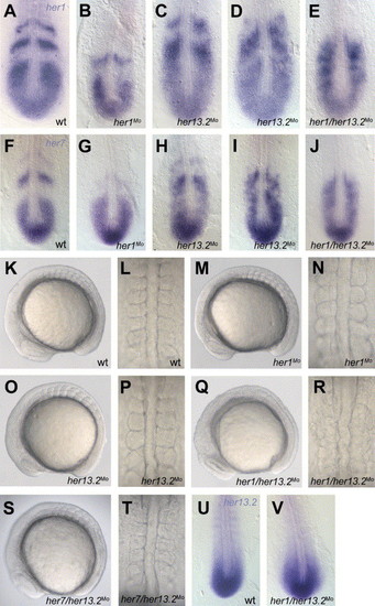
Effects of her1/her13.2 injections on somite borders and expression patterns. (A), (F) wild-type expression of her1 and her7, respectively. (B), (G) her1 and her7 expression, respectively, after her1 knockdown (0.6 mM; 2 experiments, n = 76 for each probe, 92.11% affected for her1, 94.74% affected for her7). her13.2Mo injection (0.6 mM; 2 experiments; n = 53 for her1, n = 43 for her7) leads in 73.58% of the embryos to a slightly altered her1 expression (C) and in 81.4% of the embryos to the same alteration in her7 expression (H). 9.43% of her1 stained embryos and 18.6% of her7 stained embryos show a full disruption of the her1 (D) and the her7 (I) expression, respectively. A full disruption of her1 expression (E) and her7 expression (J) is observed in her1/her13.2 double morphants (0.6 mM her1Mo + 0.6 mM her13.2Mo; 2 experiments, n = 85, 94.11% affected for her1, 95.29% affected for her7). (K), (L) somite morphology in wild-type embryos, (M), (N) in her1 morphants (0.6 mM; 2 experiments, n = 56, all embryos show almost wild-type morphology with slight border defects up to somite 3), (O), (P) in her13.2 morphants (0.6 mM; 2 experiments, n = 52, all embryos show wild-type morphology), (Q), (R) in her1/her13.2 double morphants (0.6 mM her1Mo + 0.6 mM her13.2Mo; 2 experiments, n = 92, 97.83% affected) and (S), (T) in her7/her13.2 morphants (0.6 mM her7Mo + 0.6 mM her13.2Mo; 2 experiments, n = 46, all embryos show wild-type morphology in the anterior somites). Note in the her1/her13.2 double morphants, the head appears slightly enlarged, with a dark lens primordium, the tailbud tip has a flattened appearance (Q), and the notochord is kinked (R) compared to the wild type (K). (U) Wild-type expression of her13.2 and (V) in her1/her13.2 morphants (0.6 mM her1Mo + 0.6 mM her13.2Mo; 2 experiments, n = 42, 92.86% affected (loss of stripes might rather be a secondary effect due to loss of somite borders)). (A–J), (U), and (V) dorsal view, flat mounted embryos, anterior to the top. (K), (M), (O), (Q), and (S) lateral view, anterior to the left. (L), (N), (P), (R), and (T) dorsal view of the anterior trunk somites, anterior to the top. |
| Genes: | |
|---|---|
| Fish: | |
| Knockdown Reagents: | |
| Anatomical Terms: | |
| Stage: | 5-9 somites |
Reprinted from Developmental Biology, 293(1), Sieger, D., Ackermann, B., Winkler, C., Tautz, D., and Gajewski, M., her1 and her13.2 are jointly required for somitic border specification along the entire axis of the fish embryo, 242-251, Copyright (2006) with permission from Elsevier. Full text @ Dev. Biol.