|
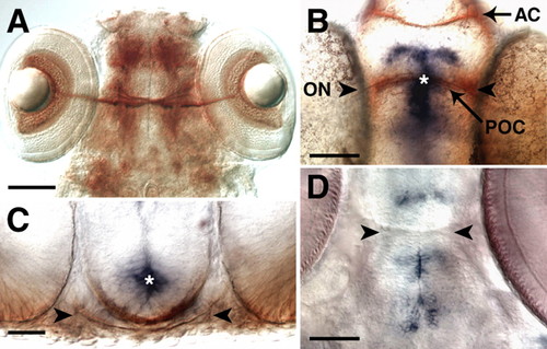
sema3d is expressed at the midline of the ventral diencephalon throughout optic pathway development. A,B,D are ventral views with anterior up; C is a cross-section with dorsal up. (A) RGC axons labeled by Zn-5 immunohistochemistry at 5 dpf form the optic chiasm in the ventral diencephalon. (B,C) sema3d in situ hybridization (blue) and α-tubulin immunohistochemistry (brown) at 36 hpf (B) and 38 hpf (C) show sema3d expression at the midline of the ventral diencephalon, anterior, posterior and dorsal to where RGC axons cross the midline. (D) sema3d expression is maintained in the diencephalon through 5 dpf. Arrowheads show the location of RGC axons, and asterisks indicate the same region of sema3d in B and C. AC, anterior commissure; ON, optic nerve; POC, postoptic commissure. Scale bars: 100 μm in A; 50 μm in B,D; 25 μm in C.
|