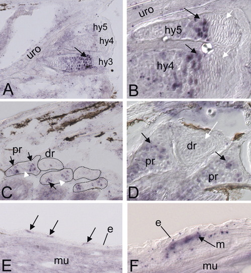
ihha expression in fin endoskeleton and scales. A-F: In situ hybridization using a ihha probe on longitudinal sections of 10-mm larvae. A,B: Caudal fin endoskeleton. A: ihha expression in the hypural of the caudal fin. B: A closer view of A reveals that the distalmost chondrocytes of the hypurals (white arrows) present a different morphology, and do not express ihha. C,D: Dorsal fin endoskeleton. C: ihha is expressed in cells located at the extremities of the proximal radial (pr, black arrows), but not in central cells (white arrowhead). D: Closer view of the dorsal fin endoskeleton showing ihha expression in chondrocytes of the proximal radials (black arrows), but not in the distal radials (white arrows). E: ihha is expressed in growing scales (arrows). Higher magnification (F) shows that ihha expression is undetectable in the epidermis and appears to be restricted in the underlying mesenchyme (arrow on F). e, epidermis; dr, distal radials; hy, hypurals; m, mesenchyme; mu, muscle; pr, proximal radials; uro, urostyle. All sections are oriented anterior to the left and dorsal to the top.
|
