
Fig. 5
- ID
- ZDB-FIG-051105-5
- Publication
- Rieger et al., 2005 - Quantum dots are powerful multipurpose vital labeling agents in zebrafish embryos
- Other Figures
- All Figure Page
- Back to All Figure Page
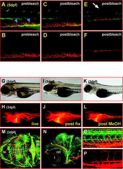
Streptavidin-conjugated QD605 are extremely photostable in vivo and withstand fixation for immunohistochemical analyses. A-F: Lateral views of the trunk of a 5-day-old zebrafish larva that has been soaked overnight in Bodipy Ceramide (green fluorescence) and was subsequently subjected to QD605 microangiography (red fluorescence). Bodipy Ceramide fluorescence in the trunk muscles and notochord diminishes after 150 consecutive 488-nm laser scans (7.9 sec each) of 3.75 mW (compare A and C), whereas the QD605 fluorescence of the reticular cells remains unaltered (compare B and D). E,F: Thus, at lower magnification, an area with bleached Bodipy Ceramide labeling can be observed (E) when compared with adjacent tissue, whereas the strength of the QD605 fluorescence appears equally strong throughout the trunk (F, see also Supplementary Movie 5 for bleaching dynamics). G-L: Lateral views of the head of a 3-day-old zebrafish larva that has been subjected to streptavidin-conjugated QD605 microangiography; strong labeling of the vasculature can be observed in the living specimen (G,H) that remains after fixation overnight in 4% paraformaldehyde in phosphate buffered saline containing 0.1%Tween 20 (I,J) and appears still unaffected after storage in methanol at -20°C and subsequent rehydration (K,L). M-P: Such embryos were subsequently subjected to immunohistochemistry using an antibody against acetylated tubulin to mark axon tracts of differentiating neurons in the brain (M, dorsal view, maximum intensity projection of 49 sections each 2 μm apart; N, lateral view, maximum intensity projection of 29 sections each 2 μm apart; images were recorded using the Meta channel of a Zeiss LSM510 laser scanning microscope, fluorescence was recorded from 499 to 670 nm in steps of 10.7-nm intervals, the emission of both fluorophores was subsequently separated by linear unmixing) and the trunk (O, lateral view, maximum intensity projection of 57 sections each 2 m apart; P, QD605 emission only, to highlight the blood vessel network within the trunk; images were recorded using the Meta channel of a Zeiss LSM510 laser scanning microscope, fluorescence was recorded from 499-670 nm in steps of 10.7-nm intervals, the emission of both fluorophores was subsequently separated by linear unmixing) for double-labeling purposes. This finding shows that blood vessels obey the segmented pattern of the brain and trunk. cb, cerebellum; l, lens; ot, optic tectum; ret, retina; rh, rhombencephalon; te, telencephalon; dpf, days postfertilization. |