
FIGURE
Fig. 6
- ID
- ZDB-FIG-050906-10
- Publication
- Chen et al., 2005 - A unique role for 6-O sulfation modification in zebrafish vascular development
- Other Figures
- All Figure Page
- Back to All Figure Page
Fig. 6
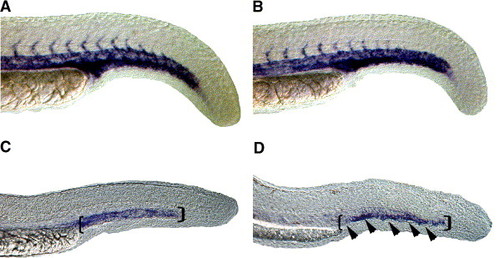
Expression of late endothelial markers in the region of forming venous plexus is reduced in HS6ST-2 morphants. Expression of flk-1 as shown by whole-mount in situ hybridization in a wild-type embryo (A) and a HS6ST-2 MO-injected embryo (B) at 24 hpf. Normal expression of flk-1 is observed in HS6ST-2 MO-injected embryos at 24 hpf (n = 19). However, at 30 hpf, the expression domain of the late endothelial marker, tie-1 (D; 81% ± 8%, n = 40, ±SEM) is reduced in the region of caudal vein (area indicated by brackets and arrowheads) in HS6ST-2 MO-injected embryos compared to wild-type embryos (C). |
Expression Data
| Genes: | |
|---|---|
| Fish: | |
| Knockdown Reagent: | |
| Anatomical Terms: | |
| Stage Range: | Prim-5 to Prim-15 |
Expression Detail
Antibody Labeling
Phenotype Data
Phenotype Detail
Acknowledgments
This image is the copyrighted work of the attributed author or publisher, and
ZFIN has permission only to display this image to its users.
Additional permissions should be obtained from the applicable author or publisher of the image.
Reprinted from Developmental Biology, 284(2), Chen, E., Stringer, S.E., Rusch, M.A., Selleck, S.B., and Ekker, S.C., A unique role for 6-O sulfation modification in zebrafish vascular development, 364-376, Copyright (2005) with permission from Elsevier. Full text @ Dev. Biol.