- Title
-
Differential induction of four msx homeobox genes during fin development and regeneration in zebrafish
- Authors
- Akimenko, M.A., Johnson, S.L., Westerfield, M., and Ekker, M.
- Source
- Full text @ Development
|
Expression of four msx genes in pectoral fin buds and their primordia. (A) Cells on the surface of the yolk sac in the primordia of the pectoral fin buds (arrows) express msxB at 24 h. The hybridization signal indicated by the arrowhead corresponds to msxB expression by cells of the dorsal hindbrain and spinal cord (Ekker et al., unpublished observations). (BE) Dissected pectoral fin buds from 48 h zebrafish embryos. Anterior is to the left, distal to the top. Cells in the AER as well as cells that occupy a more proximal position next to the AER express msxA, msxB and msxD (B, C and E, respectively). There are also weak hybridization signals for both msxA and msxD throughout the fin bud. (D) Expression of msxC at 48 h is restricted to cells next to the AER and is more intense along the anterior edge. Scale bar: 20 μm in (B,C); 40 μm in (D,E). |
|
Cells in the median fin fold express msx genes. (A) Transverse section through the tailbud of a 16 h zebrafish embryo. The dorsal side is to the top. Cells on the median edge of the tail bud express msxB. (B) At 24 h, transcripts of the msxB genes are uniformly distributed along the median fin fold. At 36 h, the most distal cells of the median fin fold and cells that occupy an immediately more proximal position express msxA, msxB and msxD (E, F and H, respectively). In contrast, MsxC expression at 36 h is restricted to cells of the median fin fold that occupy a position immediately proximal to the most distal cells (G). (C,D) Transverse sections through the tail of 48 h zebrafish embryos: msxC expression is confined to the mesenchymal cells of the fin fold (C), msxD transcripts are also found in the mesenchymal cells as well as in the ectodermal cells (D). The arrows indicate cells of the dorsal spinal cord and overlying ectoderm that also express msxB (B,F) and msxC (G). Cells in or around the vent also express all four msx genes (arrowheads in E-H). Embryos in (E-G) were treated with 0.2 mM phenylthiocarbamide to inhibit pigmentation. nk, neural keel; n, notochord. Scale bar: 20 μm in (A,C,D), 40 μm in (B,E-H). EXPRESSION / LABELING:
|
|
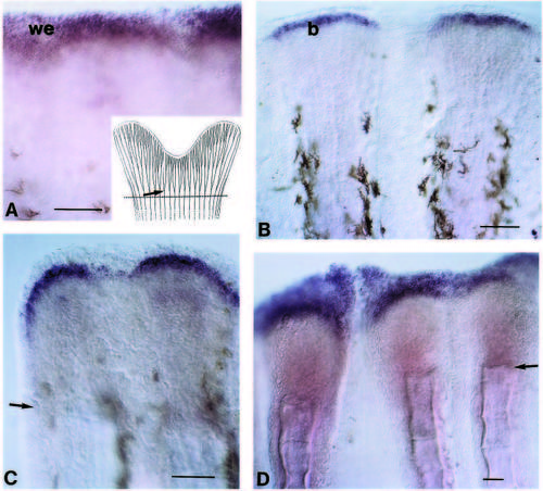
Induction of msx gene expression during regeneration of the caudal fin. Blastema cells at the tip of each fin ray express msxB (B) and msxC (C). Cells overlying the blastema express msxA and msxD (A,D). Gene expression was determined 3 d after amputation. The plane of amputation is indicated by an arrow in C-D. The inset in A is the schematic representation of the skeleton of a zebrafish caudal fin. The standard plane of amputation is indicated by a dashed line and corresponds to a level proximal to the first branch point of the lepidotrichia (arrow). Scale bar: 40 μm. EXPRESSION / LABELING:
|
|
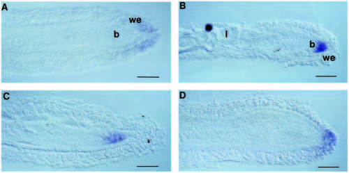
Cell-specific expression of the msx genes during regeneration of the caudal fin. Sections along the proximodistal axis of fins hybridized with probes for msxA (A), msxB (B), msxC (C) and msxD (D).Gene expression was determined 3 d after amputation. b, blastema; l, lepidotrichia; we, wound epidermis. Scale bar: 20 μm. EXPRESSION / LABELING:
|
|
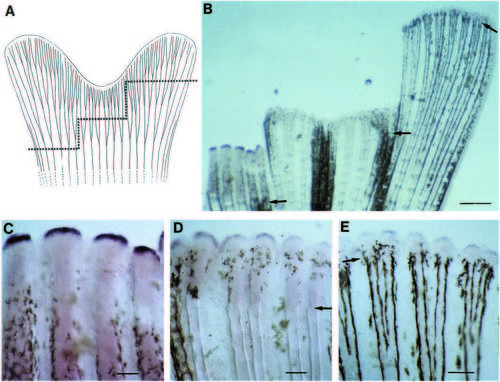
Differential expression of the msxB gene along the proximodistal axis of the regenerating fin. (A) Amputation of a single zebrafish caudal fin at three different levels along the proximodistal axis. The skeletal elements of the fin are shown as in Fig. 5A. (B) msxB expression in blastema cells of regenerating rays of a fin which was partly amputated at three different levels. (C-E) Higher magnification photographs of the fin shown in B; (C) proximal cut; (D) intermediate cut; (E) distal cut. Gene expression was determined 4.5 d after amputation. The plane of amputation is indicated by a dashed line in A and by arrows in B,D,E. Scale bar: 500 μm in B; 100 μm in C-E. EXPRESSION / LABELING:
|

Unillustrated author statements EXPRESSION / LABELING:
|


