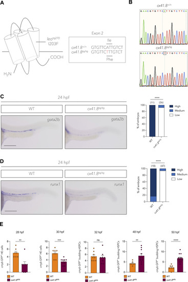
The I203F mutation in Cx41.8 results in a defect in haemogenic endothelium induction and HSPC specification. (A) The leotq270 (cx41.8tq/tq) mutant possesses an I203F change in the fourth transmembrane domain. (B) Sanger sequencing shows an A-to-T base change in the cx41.8tq/tq mutant. (C) In situ hybridisation and quantification of gata2b in cx41.8tq/tq mutants and controls at 24 hpf. (D) In situ hybridisation and quantification of runx1 in cx41.8tq/tq mutants and controls at 24 hpf. (E) Quantification of cmyb:GFP+ haemogenic endothelial (HE) cells and HSPCs in cx41.8tq/tq and control embryos between 28 and 50 hpf. Statistical significance was calculated using either a Chi-squared test (C and D) or an unpaired t-test (E). *P<0.05, **P<0.01, ***P<0.001, ****P<0.0001. Scale bars: 200 μm (C and D). Created in BioRender by Petzold, T., 2025. https://BioRender.com/97nw7ib. This figure was sublicensed under CC-BY 4.0 terms.
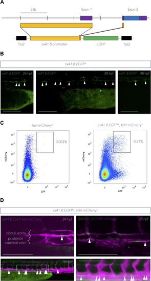
cx41.8 is expressed in presumptive haemogenic endothelial cells of the dorsal aorta. (A) Design of the cx41.8:EGFP plasmid. The upper line indicates the cx41.8 locus structure. The lower line indicates the construct design. Purple boxes indicate the cx41.8 exons; blue boxes indicate the open reading frame; yellow boxes indicate the 4.5 kb sequence upstream of the cx41.8 start codon; black boxes indicate the transposon sequences and the green box indicates the EGFP coding sequence. (B) cx41.8:EGFP expression in the presumptive floor of the aorta at 24 and 28 hpf and in presumptive budding HSPCs (48 hpf). White arrowheads denote presumptive haemogenic endothelial cells in the floor of the aorta and budding HSPCs (48 hpf). (C) Flow cytometry analysis of double-positive cells in 48 hpf kdrl:mCherry+ or cx41.8:EGFP+; kdrl:mCherry+ embryos. (D) Expression of cx41.8:EGFP and kdrl:mCherry from 24-48 hpf. White arrowheads denote cx41.8:EGFP and kdrl:mCherry double-positive endothelial cells in the floor of the dorsal aorta. Scale bars: 100 μm (B and D).
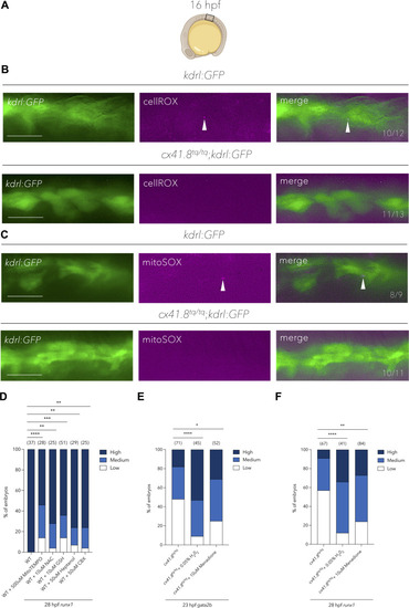
Mitochondrial-derived ROS production in endothelial cells is required for haemogenic endothelium induction and the specification of HSPCs. (A) Schematic showing the region of 16 hpf embryos which was analysed by fluorescence microscopy in B and C. (B) Fluorescence microscopy images of total cellular ROS detection in kdrl:GFP+ or cx41.8tq/tq;kdrl:GFP+ embryos. White arrowheads denote the presence of ROS in endothelial cells. Numbers indicate the ratio of embryos with the respective phenotype. (C) Fluorescence microscopy images of mitochondrial-derived ROS detection in kdrl:GFP+ or cx41.8tq/tq;kdrl:GFP+ embryos. White arrowheads denote mitochondrial-derived ROS in endothelial cells. Numbers indicate the ratio of embryos with the respective phenotype. (D) Quantification of aortic runx1 signal (in situ hybridisation) at 28 hpf in WT control embryos and those treated with MitoTEMPO, NAC, GSH, heptanol or CBX. (E) Quantification of aortic gata2b signal (in situ hybridisation) at 23 hpf in cx41.8tq/tq control embryos and cx41.8tq/tq embryos treated with H202 or menadione. (F) Quantification of aortic runx1 signal (in situ hybridisation) at 28 hpf in cx41.8tq/tq control embryos and cx41.8tq/tq embryos supplemented with H202 and menadione. Statistical significance was calculated using a Chi-squared test. *P<0.05, **P<0.01, ***P<0.001, ****P<0.0001. Scale bars: 25 μm (B and C). Created in BioRender by Petzold, T., 2025. https://BioRender.com/n88jg67. This figure was sublicensed under CC-BY 4.0 terms.
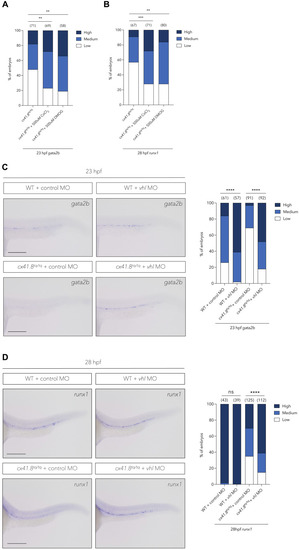
Stabilisation of Hif1/2α rescues haemogenic endothelium induction and HSPC specification in cx41.8tq/tq mutants. (A) Quantification of aortic gata2b signal (in situ hybridisation) at 23 hpf in cx41.8tq/tq control embryos and cx41.8tq/tq embryos supplemented with CoCl2 or DMOG. (B) Quantification of aortic runx1 signal (in situ hybridisation) at 28 hpf in cx41.8tq/tq control embryos and cx41.8tq/tq embryos treated with CoCl2 or DMOG. (C) In situ hybridisation and quantification of gata2b at 23 hpf in WT control embryos and cx41.8tq/tq embryos injected with either control- or vhl-MO. (D) In situ hybridisation and quantification of runx1 at 28 hpf in control-MO or vhl-MO injected wild-type or cx41.8tq/tq mutant embryos. Statistical significance was calculated using a Chi-squared test. *P<0.05, **P<0.01, ***P<0.001, ****P<0.0001. Scale bars: C, 100 μm, D, 200 μm.
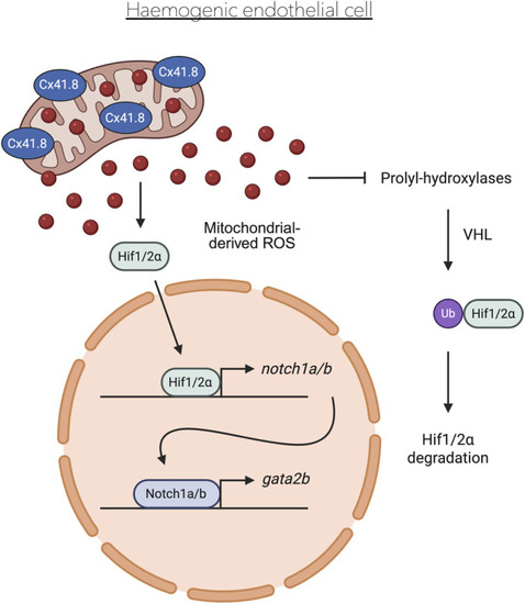
Proposed model of the role of Cx41.8 in HSPC specification. Cx41.8 localises to the mitochondria in haemogenic endothelial cells, allowing mitochondrial ROS production which stabilises Hif1/2α, which in turn induces gata2b expression via Notch1a/b signalling. Created in BioRender by Petzold, T., 2025. https://BioRender.com/konhezh. This figure was sublicensed under CC-BY 4.0 terms.
Acknowledgments
This image is the copyrighted work of the attributed author or publisher, and
ZFIN has permission only to display this image to its users.
Additional permissions should be obtained from the applicable author or publisher of the image.
Full text @ Biol. Open
Your Input Welcome
Thank you for submitting comments. Your input has been emailed to ZFIN curators who may contact you if
additional information is required.
Oops. Something went wrong. Please try again later.