FIGURE SUMMARY
Title

The role of 25-hydroxycholesterol in the pathophysiology of brain vessel dysfunction associated with infection and cholesterol dysregulation

Authors
Tapia, V.S., Withers, S.E., Zhou, R., Bennington, A., Hoyle, C., Hedley, F., El Khouja, A., Luka, N., Massimo, M., Crilly, S., Long, K.R., Lawrence, C.B., Kasher, P.R.
Source
Full text @ Dis. Model. Mech.

SARS-CoV-2 spike protein induces ch25h upregulation prior to spontaneous brain bleeding in zebrafish larvae. (A) Representative images of control [intracerebral haemorrhage (ICH)] and haemorrhagic (ICH+) Tg(fli1:EGFP)/Tg(gata1a:DsRed) 3 days post-fertilisation (dpf) larvae, 24 h after SARS-CoV-2 spike protein (spike) injection into the hindbrain (0.25 mg ml−1, 2 nl). Red indicates erythrocytes (gata1a+) and cyan indicates endothelial cells (fli1+). Dashed lines indicate the brain area. Scale bars: 250 µm. (B) Time course of ICH+ frequency in Tg(fli:EGFP)/Tg(gata1a:DsRed) larvae that were uninjected, injected with bovine serum albumin (BSA) control, injected with pre-heated spike at 80oC for 30 min (spike-80) or injected with spike conditions (n=4, 11-14 embryos per experiment). (C) Haematoma size (Gata+ area in brain region) in larvae 24 h post-injection with BSA or spike. Individual embryos are indicated as dots (n=146-147, three independent experiments). (D,E) Gene expression was analysed in larval heads 8 and 14 h after BSA or spike injections, for ch25h (D), ch25hl1.1, ch25hl2 and ch25hl3 (E) (30 larval heads pooled per replicate). Data are mean±s.d. (B,C,E) or median±interquartile range (IQR) (C). ns, non-significant; *P<0.05; **P<0.01; determined by repeated measures ANOVA with Dunnett's post-hoc test compared to uninjected (B), Mann–Whitney test (C) or randomised block two-way ANOVA with Sidak's post hoc analysis compared to BSA (D,E).

CH25H expression is associated with human foetal SARS-CoV-2-associated brain microbleeds. (A) Representative images of microbleeds found in human foetal cortex by Haematoxylin and Eosin staining. Bleeds were classified into small, medium and large sizes according to area. Scale bar size is shown in each image. (B,C) Samples were categorised according to a bleeding score, based on bleed size and total bleed density. Control samples were classified as score 0, and haemorrhagic samples were classified as scores 1 and 2. Density of small, medium and large bleeds (B), and total bleed density (C) is shown for samples with bleeding scores of 0 to 2. (D) Representative images of CH25H staining (pink) and bleeds (yellow) from non-haemorrhagic or haemorrhagic samples, proximal and distal to bleeds. Scale bars: 30 µm. (E) CH25H+ cell density was quantified in samples with bleeding scores of 0 to 2. Data are mean±s.e.m. ns, non-significant; **P<0.01; ***P<0.001; determined by two-way ANOVA with Tukey's post hoc analysis multiple comparisons between scores (B), or one-way ANOVA with Kruskal–Wallis post hoc test compared to score 0 (C,E).

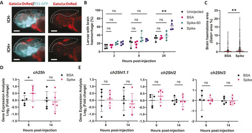
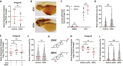
25HC worsens brain haemorrhage expansion in a statin-induced ICH zebrafish model. (A) hmgcrb expression in 2 days post-fertilisation (dpf) wild-type (WT) zebrafish larvae incubated with 25-hydroxycholesterol (25HC; 25 μM, 24 h) (15 embryos pooled per replicate). (B-D) WT larvae were incubated in Atorvastatin (ATV; 1 µM) at 28 h post-fertilisation (hpf) and intravenously injected with 25HC (1 nl, 5 µM) at 32-36 hpf. The next day, larvae were stained with o-Dianisidine. Representative images of larvae without (ICH) and with (ICH+) brain haemorrhage are shown (B). Dotted lines indicate the brain area. Scale bars: 150 µm. ICH+ frequency per experiment (C) and brain haematoma area per larvae (D) were quantified. Individual embryos are indicated as dots (n=128 embryos, five independent experiments). (E) hmgcrb expression in 2 dpf WT larvae incubated with 25HC (25 µM) and ATV (1 µM) for 24 h (15 embryos pooled per replicate). (F) bbh zebrafish larvae were injected with 25HC (1 nl, 5 µM) at 32-36 hpf. After 24 h, larvae were stained with o-Dianisidine and brain haematoma area was quantified. Individual embryos are indicated as dots (n=69-82 embryos, three independent experiments). (G) Comparison of 25HC and 4β-hydroxycholesterol (4βHC) structures. (H) Expression of hmgcrb in 2 dpf WT larvae incubated with 4βHC or 25HC (25 µM, 24 h) (15 embryos pooled per replicate). (I) WT larvae were incubated in ATV (1 µM) at 28 hpf and injected with 4βHC or 25HC (1 nl, 2.5 µM) at 32-36 hpf. The next day, larvae were stained with o-Dianisidine and haematoma area was quantified. Individual embryos are indicated as dots (n=87-93 embryos, three independent experiments). Data expressed as mean±s.d. (A,C,E,H) or median±IQR (D,F,I). ns, nonsignificant; *P<0.05; **P<0.01; determined by paired two-tailed t-test (A), randomised block two-way ANOVA with Sidak's post-hoc test compared to control (C), Mann–Whitney test (D,F), randomised block one-way ANOVA with Dunnett's post-hoc test compared to control (E,H), or Kruskal–Wallis test with Dunn's post-hoc test compared to control (I).
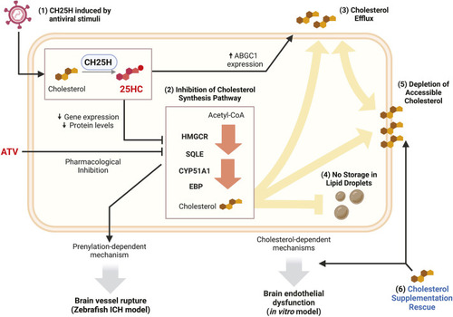
25HC remodels cholesterol metabolism in human brain endothelial cells. (A) Cellular cholesterol remodelling induced by 25HC. 25HC inhibits cholesterol synthesis (1), promotes cholesterol efflux (2), and induces the internalisation and storage of cholesterol in the form of lipid droplets (3). These changes lead to the depletion of plasma membrane-accessible cholesterol (4), which can be rescued with cholesterol supplementation (5). (B,C) Expression of HMGCR, SQLE, CYP51A1, EBP (B) and ABCG1 (C) genes in hCMEC/D3 cells after 25HC treatment (5 μM, 4 and 24 h). (D) hCMEC/D3 cells were loaded with fluorescent cholesterol (1 h) before 25HC treatment (5 μM, 16 h). Cholesterol efflux in fresh medium (for 4 h) was measured by fluorescence. (E,F) hCMEC/D3 cells were pre-treated with 25HC (5 μM, 0 to 24 h) before incubation with streptolysin O (SLO). Membrane permeability was measured by To-Pro-3+ uptake (red signal) before and after SLO; representative images (E) and quantification (F) are shown. Scale bars: 37.5 µm. (G) Permeability of hCMEC/D3 cells, pre-treated with 25HC (5 μM, 6 h) and then with soluble cholesterol (80 μM, 1 h), after SLO incubation. Data expressed as mean±s.d. ns, nonsignificant; *P<0.05; **P<0.01; ***P<0.001; ****P<0.0001; determined by paired two-tailed t-test (D), randomised block one-way ANOVA with Dunnett's post hoc test compared to 0 µM (F), or randomised block two-way ANOVA with Sidak's post-hoc test compared to control (B,C,G).

25HC dysregulates the function of human brain endothelial cells. (A) Permeability of fluorescein-conjugated dextran 70 kDa (FD70) in hCMEC/D3 cell monolayer pre-treated with different concentrations of 25HC (0-5 μM, 24 h). (B,C) hCMEC/D3 cell migration in a scratch assay; cells were pre-treated with different concentrations of 25HC (0-5 μM, 24 h before scratch). Representative images are shown for cells 0 and 24 h after scratch (B; 5 μM 25HC). Blue lines show the initial scratched area. Scale bars: 100 µM. Migration was quantified as relative wound density (C; n=10). (D) FD70 permeability was analysed in hCMEC/D3 cells treated with 25HC (5 μM, 24 h) and ATV (1 μM, 24 h). (E) Cell migration at 24 h post-scratch of hCMEC/D3 cells pre-treated with 25HC (5 μM, 24 h) before scratch and treated with ATV (1 µM, 24 h) after scratch. (F) FD70 permeability was analysed in hCMEC/D3 cells pre-treated with 25HC (5 μM, 14 h) and then supplemented with soluble cholesterol (80 µM, 1 h). (G) Cell migration at 24 h post-scratch of hCMEC/D3 cells pre-treated with 25HC (5 μM, 24 h) and then supplemented with cholesterol (80 µM, 2 h) before scratch. Data expressed as mean±s.d. ns, nonsignificant; *P<0.05; **P<0.01; ***P<0.001; ****P<0.0001; determined by randomised block one-way ANOVA with Dunnett's post-hoc test compared to 0 µM (A), matched measures two-way ANOVA with Dunnett's post-hoc test compared to 0 µM (C), or randomised block two-way ANOVA with Sidak's post-hoc test compared to control (D-G).

25HC remodels cholesterol metabolism and function of brain endothelial cells. (1) CH25H upregulation was detected in SARS-CoV-2-associated ICH in zebrafish and human foetal brain tissue, as well as in hCMEC/D3 cells exposed to antiviral stimuli [poly(I:C) and IFNβ]. (2) 25HC treatment induced the downregulation of cholesterol synthesis enzymes at both mRNA and protein levels. In zebrafish and hCMEC/D3 cells, this downregulation had an additive effect when combined with pharmacological inhibition of HMGCR by ATV, leading to brain vessel rupture (ICH) in zebrafish and decreased barrier function and cell migration in hCMEC/D3 cells. (3) 25HC treatment in hCMEC/D3 cells also increased cholesterol efflux, which was associated with upregulation of ABCG1. (4) Internalisation of cholesterol into lipid droplets was not observed in 25HC-treated hCMEC/D3 cells. (5) The changes in cholesterol synthesis and efflux in hCMEC/D3 cells were associated with the depletion of plasma membrane-accessible cholesterol. (6) Cholesterol supplementation rescued the levels of accessible cholesterol and mitigated the decrease in barrier function and cell migration induced by 25HC in hCMEC/D3 cells.

Acknowledgments
This image is the copyrighted work of the attributed author or publisher, and ZFIN has permission only to display this image to its users. Additional permissions should be obtained from the applicable author or publisher of the image. Full text @ Dis. Model. Mech.