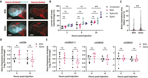
SARS-CoV-2 spike protein induces ch25h upregulation prior to spontaneous brain bleeding in zebrafish larvae. (A) Representative images of control [intracerebral haemorrhage (ICH)−] and haemorrhagic (ICH+) Tg(fli1:EGFP)/Tg(gata1a:DsRed) 3 days post-fertilisation (dpf) larvae, 24 h after SARS-CoV-2 spike protein (spike) injection into the hindbrain (0.25 mg ml−1, 2 nl). Red indicates erythrocytes (gata1a+) and cyan indicates endothelial cells (fli1+). Dashed lines indicate the brain area. Scale bars: 250 µm. (B) Time course of ICH+ frequency in Tg(fli:EGFP)/Tg(gata1a:DsRed) larvae that were uninjected, injected with bovine serum albumin (BSA) control, injected with pre-heated spike at 80oC for 30 min (spike-80) or injected with spike conditions (n=4, 11-14 embryos per experiment). (C) Haematoma size (Gata+ area in brain region) in larvae 24 h post-injection with BSA or spike. Individual embryos are indicated as dots (n=146-147, three independent experiments). (D,E) Gene expression was analysed in larval heads 8 and 14 h after BSA or spike injections, for ch25h (D), ch25hl1.1, ch25hl2 and ch25hl3 (E) (30 larval heads pooled per replicate). Data are mean±s.d. (B,C,E) or median±interquartile range (IQR) (C). ns, non-significant; *P<0.05; **P<0.01; determined by repeated measures ANOVA with Dunnett's post-hoc test compared to uninjected (B), Mann–Whitney test (C) or randomised block two-way ANOVA with Sidak's post hoc analysis compared to BSA (D,E).
|
