FIGURE SUMMARY
Title

Endogenous acrolein accumulation in akr7a3 mutants causes microvascular dysfunction due to increased arachidonic acid metabolism

Authors
Zhang, X., Gschwind, J., Erben, V., Bennewitz, K., Li, X., Sticht, C., Poschet, G., Hausser, I., Fleming, T., Szendroedi, J., Nawroth, P.P., Kroll, J.
Source
Full text @ Redox Biol.

Endogenous ACR was accumulated in akr7a3−/− mutant zebrafish. (A–D) ELISA determination indicated that ACR was significantly increased in 96 hpf old akr7a3−/− larvae (A) and in adult akr7a3−/− liver (B), akr7a3−/− eye (C), and akr7a3−/− kidney (D). Each data point indicates 47–50 larvae per clutch or one adult zebrafish organ, n = 5–8. (E–H) Representative RCS quantification by mass spectrometry or ELISA. The concentration of glyoxal (E), methylglyoxal (F), 3DG (G), and 4-HNE (H) remained unaltered in akr7a3−/− larvae. Each data point indicates 47–50 larvae per clutch, n = 6. (I–K) Quantification of GSH and GSSG revealed a significant decrease in GSH in 5 dpf old akr7a3−/− larvae (I), while GSSG (J) levels and the GSH/GSSG ratio (K) remained unchanged. Data represent 50 larvae per clutch, n = 5. The bars indicate mean ± SD values. Statistical analysis was performed by Student's t-test. ACR, acrolein; 3DG, 3-Deoxyglucosone; 4-Hydroxynonenal, 4-HNE; GSH, glutathione; GSSG, glutathione disulfide.

Increased hyaloid vessel diameter in larvae of akr7a3−/− eyes. (A–C) Representative confocal images and quantification of hyaloid vasculature in larvae at 5dpf. No significant difference was observed in branches among akr7a3+/+ and akr7a3−/− mutant larvae (B), however the diameter of hyaloid blood vessels was significantly increased in akr7a3−/− mutants (C), white scale bar = 30 μm, n = 40/28, red arrows indicate branch points. (D–G) Representative confocal images and quantification of adult retinal vasculature. The vascular branches (E), sprouts (F), and vascular density (G) were not altered between adult akr7a3+/+ and akr7a3−/− zebrafish. Yellow arrows are branch points, red circles are sprouts. Red frame, high density area; Blue frame, low density area; Yellow frame, middle density area; Upper white scale bar = 350 μm, lower white scale bar = 100 μm, n = 7/8. Each data point represents vasculature in a 350 μm2 area in the high-density region of the retina. Statistical analysis was performed by Student's t-test, The bars indicate mean ± SD values; dpf, days post fertilization.

Increased retinal vessel diameters and GBM thickening in adult akr7a3−/− zebrafish. (A–D) Representative hematoxylin staining and quantification of retinal vasculature. The retinal blood vessel diameters of akr7a3−/− eyes were significantly increased (B). The number of MC (C) and EC (D) was also increased. n = 40. Blue arrows, EC; green arrows, MC; Red arrows, RBC; Black scale bar = 100 μm. (E,F) Representative EM images and GBM quantification showed a significant increase in GBM thickness in adult akr7a3−/− kidneys. n = 15/18. Black arrow, normal GBM of adult akr7a3+/+ zebrafish, red arrow, thickened GBM of akr7a3−/− zebrafish. Scale bar = 10 μm (left) and 2 μm (right), respectively. The bars indicate mean ± SD values. Statistical analysis was performed by Student's t-test. EC, endothelial cells; MC, mural cell; RBC, red blood cell; EM, electron microscopy; GBM, glomerular basement membrane.

Exogenous ACR induced dilation of hyaloid blood vessels and pronephric nephron alterations in zebrafish. (A–B) Representative confocal images and quantification of hyaloid vasculature in larvae at 5dpf. A significant increase in diameter of hyaloid vasculature was observed in akr7a3+/+ larvae incubated with 10 μM ACR. n = 27–30, white scale bar = 30 μm. (C) Fluorescence microscopy images of pronephric nephrons at 48hpf. Larvae of akr7a3+/+ were incubated with ACR, ACR&CAR, or equivalent volume of DMSO as indicated, white scale bar = 50 μm. (D–F) Quantification of pronephric nephron indicated unchanged glomerular length (D) and glomerular width (E). However, neck length (F) was significantly reduced after ACR incubation compared to control group, while the reduced neck length was rescued in ACR&CAR co-incubation group. n = 9/10. (G–H) Expression levels of nephrin (G) and podocin (H) were significantly reduced in adult akr7a3−/− kidneys, indicating a marked disruption of the glomerular filtration barrier in the absence of Akr7a3. n = 5. For statistical analysis, one-way ANOVA was used for comparisons among multiple groups, while the Student's t-test was applied for comparisons between two groups. Data are presented as mean ± SD. CAR, l-Carnosine; DMSO, Dimethyl sulfoxide.

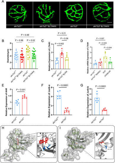
Altered arachidonic acid metabolism pathway in akr7a3−/− zebrafish. (A) Pathway enrichment analysis based on metabolomic data indicates loss of akr7a3 altered multiple metabolism pathways, most of which are involved in lipid metabolism. (B) Clustering analysis of RNA-seq disclosed that the gene expression profiles were significantly altered in adult eyes between akr7a3−/− and akr7a3+/+ zebrafish. (C). GO/KEGG enrichment analysis of RNA-seq confirmed the alteration of lipid metabolic pathway, however the RNA-seq data also disclosed considerable metabolic changes of amino acids, glucose, and antioxidants between akr7a3−/− and akr7a3+/+ zebrafish eyes as indicated. (D) Brief illustration of arachidonic acid metabolism pathway. Arachidonic acid is released from membrane phospholipids by the action of enzymes like PLA2/PLC and can then undergo various metabolic pathways and produces several bioactive lipid mediators include PGs, EETs, DHETs, and LTs. (E–H) Based on pathway enrichment analysis, expression of downstream targets gene including LTA4H, sEH, COX-2, and CYP2 was analyzed. Quantification of mRNA expression indicated an increase of LTA4H and sEH (E,H), a decrease of COX-2 (F), and an unchanged CYP2 in akr7a3−/− mutants (G). n = 5 clutches with 30 larvae and n = 5 the adult liver and eye group. Student's t-test was applied for statistical analysis. PLA2/PLC, Phospholipase A2/C; LTA4H, Leukotriene A4 hydrolase; sEH, soluble epoxide hydrolase; COX-2, Cyclooxygenase-2; CYP2, Cytochrome P450 2. PGs, Prostaglandins; EETs, Epoxyeicosatrienoic acids; DHETs, Dihydroxyeicosatrienoic acids; LTs, Leukotrienes; HETEs, Hydroxyeicosatetraenoic acids; PGH2 Prostaglandin H2; ALOX, Arachidonate 5-lipoxygenase; LXs, Lipoxins; LTB4/C4/D4/E4, Leukotriene B4/C4/D4/E4.

LTA4H was upregulated by ACR and induced vascular alteration in akr7a3−/− zebrafish. (A–B) Representative confocal images and quantification of hyaloid blood vessel diameters in larvae at 5dpf. The increased diameter of hyaloid vasculature in akr7a3−/− zebrafish was significantly reduced after LTA4Hi incubation. White scale bar = 30 μm, n = 18–20. (C–D) RT-qPCR analysis of LTA4H expression. LTA4H was significantly increased in akr7a3−/− zebrafish larvae. Incubation with CAR reduced the expression of LTA4H non-significantly. After incubating akr7a3+/+ zebrafish larvae with ACR, an increase in LTA4H expression was observed. However, co-incubation with ACR and CAR was able to inhibit the elevated expression of LTA4H. n = 6 clutches with 30 larvae. (E–G) RT-qPCR analysis data revealed an increased expression of AhR (E), while Junb-a (F) and Junb-b (G) exhibited decreased expression. n = 6 clutches with 30 larvae. (H–I) Docking analysis showed ACR (I) shared same binding pocket with Indirubin (H). ACR formed a hydrogen bond with the amino residue SER336 of AhR. Dash line = hydrogen bonds. For statistical analysis one-way ANOVA or Student's t-test was applied. The bars indicate values of mean ± SD. AhR, Aryl hydrocarbon receptor; Junb-a, JunB Proto-Oncogene a; Junb-b, JunB Proto-Oncogene b.

Upregulated activity of LTB4R promoted inflammatory response and vascular alteration in akr7a3−/− zebrafish. (A–D) The expression of leukotriene receptors was examined. It revealed that the expression of CysLT1R (A), CysLT2R (B), and CysLT3R (C) were not altered but most gene showed an upregulated trend in akr7a3−/− group whereas LTB4R (D) was significantly overexpressed. n = 5 clutches with 30 larvae. (E–F) Rescue experiment using antagonist for LTB4R and CysLTs: Antagonists of LTB4R (U-75032) and CysLTs (Quininib) were used to incubate akr7a3−/− larvae. Treatment with U-75032 significantly reduced the retinal vessel diameter in akr7a3−/− larvae, whereas Quininib had no significant effect on the retinal vessel diameter in these larvae. n = 20 larvae. (G–J) Representative inflammatory factors were also analyzed with RT-qPCR. IL-1β (G) and TNF-α (H) were significantly overexpressed while IL-6 (I) and M-CSF (J) were increased non-sigificantly in akr7a3−/− group. n = 5 clutches with 30 larvae.The bars indicate mean ± SD values. Statistical analysis was performed by one-way ANOVA or Student's t-test. CysLTs, Cysteinyl leukotriene receptor 1 (CysLT1R), 2 (CysLT2R), and 3 (CysLT3R); LTB4R, Leukotriene B4 receptor; IL-1β, Interleukin-1 beta; TNF-α, Tumor Necrosis Factor-alpha; M-CSF, IL-6, interleukin 6; Macrophage Colony-Stimulating Factor.

Acknowledgments
This image is the copyrighted work of the attributed author or publisher, and ZFIN has permission only to display this image to its users. Additional permissions should be obtained from the applicable author or publisher of the image. Full text @ Redox Biol.