
- Title
-
Wnt2bb signaling promotes pharyngeal chondrogenic precursor proliferation and chondrocyte maturation by activating Yap expression in zebrafish
- Authors
- Guo, X., Yang, L., Wang, Y., Yuan, M., Zhang, W., He, X., Wang, Q.
- Source
- Full text @ J. Genet. Genomics
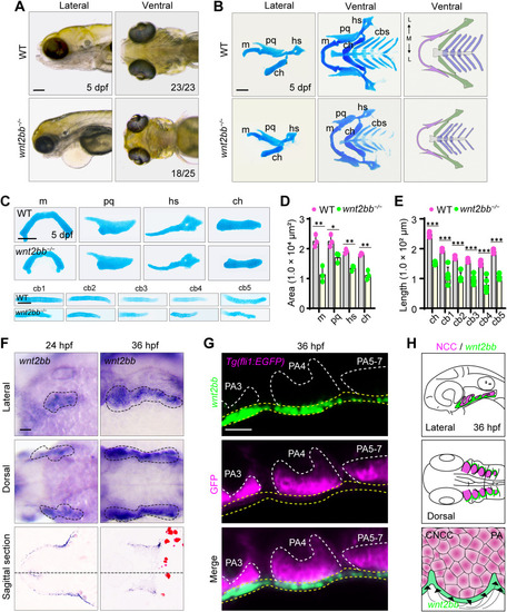
Pharyngeal ectoderm derived Wnt2bb is required for pharyngeal cartilage development. A: Bright-field images of WT and wnt2bb−/− embryos at 5 dpf. Numbers indicate the number of embryos with corresponding phenotypes out of the total number of embryos examined. B: Pharyngeal cartilages stained with Alcian blue in WT and wnt2bb−/− embryos at 5 dpf. C: Individual cartilage elements stained with Alcian blue in WT and wnt2bb−/− embryos at 5 dpf. D: Quantification of cartilage area in WT and wnt2bb−/− embryos (n = 3). E: Quantification of cartilage length in WT and wnt2bb−/− embryos (n = 5). F: WISH experiments showing the expression of wnt2bb in the pharyngeal region of WT embryos at the indicated stages. The dotted lines on the bottom panels refer to the positions of the corresponding sections. G: The expression pattern of wnt2bb in WT embryos with a Tg(fli1:EGFP) background. Embryos were subjected to a FISH experiment with a wnt2bb probe followed by immunofluorescence staining using the anti-GFP antibody. H: A schematic showing that wnt2bb is derived from the pharyngeal ectoderm and is expressed right next to the neural crest cells which subsequently differentiate into chondrocytes. ∗, P < 0.05; ∗∗, P < 0.01; ∗∗∗, P < 0.001 (student's t-test). WT, wild-type; m, Meckel's cartilage; pq, palatoquadrate; hs, hyosymplectic; ch, ceratohyal; cbs, ceratobranchial cartilages; PA, pharyngeal arch; CNCC, cranial neural crest cell; dpf, days post fertilization. Scale bars, 100 μm (A–C, F); 20 μm (G). |
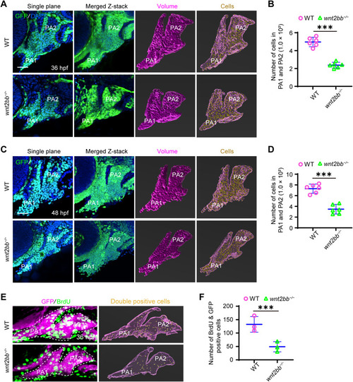
Inactivation of Wnt2bb impairs chondrocyte precursor proliferation. A and C: Single-plane and merged z-stack images of WT and wnt2bb−/− embryos with a Tg(fli1:EGFP) background at 36 hpf (A) and 48 hpf (C). Embryos were fixed and stained with anti-GFP antibodies. DAPI was used to label nuclei. Imaris software was applied to generate 3D volumes of pharyngeal arches 1 and 2, as well as virtual puncta representing cells in pharyngeal arches 1 and 2. B and D: Quantification of cell numbers in pharyngeal arches 1 and 2 of WT and wnt2bb−/− embryos at 36 hpf and 48 hpf using Imaris software (n = 6). E: Cell proliferation assays in WT and wnt2bb−/− embryos with a Tg(fli1:EGFP) background at 36 hpf (E). Virtual puncta represent BrdU incorporated cells in pharyngeal arches 1 and 2. F: Quantification of BrdU incorporated chondrogenic precursors in E (n = 3). ∗∗∗, P < 0.001 (student's t-test). WT, wild-type; PA, pharyngeal arch; hpf, hours post fertilization. Scale bar, 20 μm (A, C, E). |
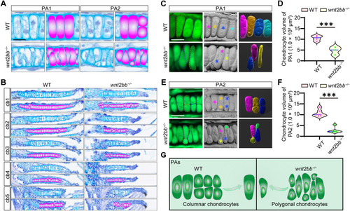
Inactivation of Wnt2bb disrupts chondrocyte maturation. A: Representative images demonstrating chondrocytes of pharyngeal arches 1 and 2 stained with Alcian blue and hematoxylin in WT and wnt2bb−/− embryos at 5 dpf. The right panels show the corresponding schematics outlining the chondrocytes. B: Representative images of ceratobranchial chondrocytes stained with Alcian blue and hematoxylin in WT and wnt2bb−/− embryos at 5 dpf. The lower panels display the corresponding schematics outlining the chondrocytes. C–F: Comparison of the size, shape, and arrangement of pharyngeal chondrocytes in WT and wnt2bb−/− embryos with a Tg(fli1:EGFP) background. 3D reconstruction was performed using Imaris software. The dotted lines and asterisks represent individual cells subjected to in-depth analysis via 3D reconstruction. The volume of each chondrocyte in pharyngeal arch 1 of WT (n = 4) and wnt2bb−/− (n = 3) embryos was quantified in (D). The volume of each chondrocyte in pharyngeal arch 2 of WT and wnt2bb−/− embryos was quantified in F (n = 6). G: An illustration demonstrating the comparison of chondrocyte size, shape, and arrangement between wild-type and wnt2bb−/− mutants. ∗∗∗, P < 0.001 (student's t-test). WT, wild-type; PA, pharyngeal arch; cb, ceratobranchial. Scale bar, 20 μm (A–C, E). |
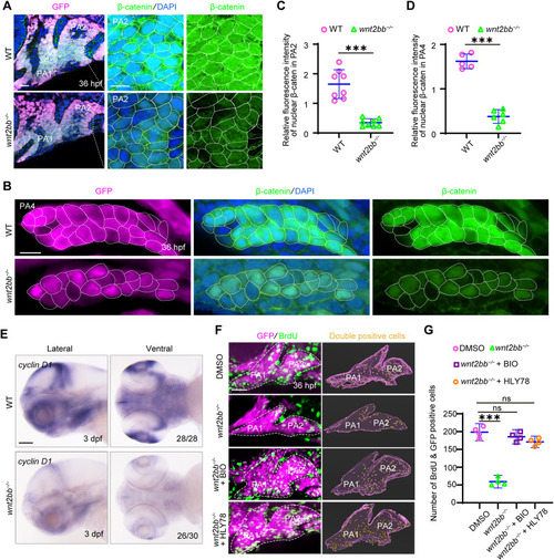
Wnt2bb promotes chondrocyte precursor proliferation by activating β-catenin dependent signaling pathway. A and B: Lateral-viewed immunofluorescence images displaying β-catenin levels in WT and wnt2bb−/− embryos with a Tg(fli1:EGFP) background at 36 hpf. Embryos were fixed and stained with anti-β-catenin and anti-GFP antibodies. Nuclei were labeled with DAPI. The dotted lines represent the magnified area of PA2 in (A). C: The relative fluorescence intensity of nuclear β-catenin in PA2 (n = 8). D: The relative fluorescence intensity of nuclear β-catenin in PA4 of WT (n = 5) and wnt2bb−/− (n = 6) embryos. E: Expression of cyclin D1 in WT and wnt2bb−/− embryos at 3 dpf. Numbers at the bottom right corner indicate the number of embryos with phenotypes shown out of the total number of embryos examined. F and G: wnt2bb−/− embryos with a Tg(fli1:EGFP) background were treated with 10 μM BIO or 10 μM HLY78 from 28 hpf, and harvested for cell proliferation assay at 36 hpf (F). Virtual puncta for BrdU incorporated cells in PA1 and PA2 were produced via Imaris software. The number of BrdU incorporated chondroprogenitors was quantified in G (n = 4). ∗∗∗, P < 0.001; ns, not significant (student's t-test). WT, wild-type; PA, pharyngeal arch; hpf, hours post fertilization; dpf, days post fertilization. Scale bars, 20 μm (A, B, F); 100 μm (E). |
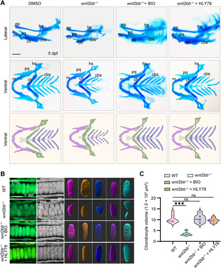
Wnt2bb controls pharyngeal chondrocyte maturation by activating β-catenin dependent signaling pathway. A and B: wnt2bb−/− embryos with a Tg(fli1:EGFP) background were treated with 10 μM BIO or 10 μM HLY78 from 28 hpf, and collected for Alcian blue staining (A) and live imaging (B) at 5 dpf, respectively. 3D reconstruction of chondrocytes (B) in ceratohyal cartilages was performed using Imaris software. The dotted lines and asterisks represent individual cells subjected to in-depth analysis via 3D reconstruction. C: The volume of individual chondrocytes in ceratohyal cartilages was quantified via Imaris (n = 7). ∗∗∗, P < 0.001; ns, not significant (student's t-test). WT, wild-type; m, Meckel's cartilage; ep, ethmoid plate; pq, palatoquadrate; hs, hyosymplectic; ch, ceratohyal; cbs, ceratobranchial cartilages; dpf, days post fertilization. Scale bars, 100 μm (A); 20 μm (B). |
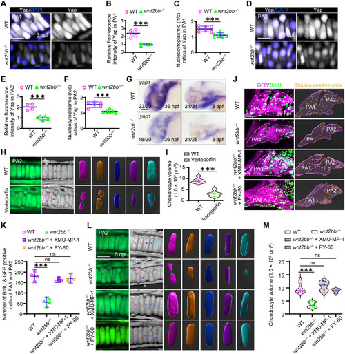
Wnt2bb signaling induces Yap expression to regulate chondrogenic precursor proliferation as well as chondrocyte maturation during pharyngeal cartilage development. A–C: Yap levels in pharyngeal arch 1 of WT and wnt2bb−/− embryos with a Tg(fli1:EGFP) background at 3 dpf. Fixed embryos were stained with anti-YAP1 and anti-GFP antibodies. Nuclei were labeled with DAPI. The relative fluorescence intensity of Yap in PA1 was quantified in B (n = 5). The nucleocytoplasmic (n/c) ratio of Yap in PA1 was shown in C (n = 6). D–F: Yap levels in PA2 at 3 dpf. The relative fluorescence intensity of Yap in PA2 of WT (n = 4) and wnt2bb−/− (n = 5) embryos was quantified in (E). The nucleocytoplasmic (n/c) ratio of Yap in PA2 was displayed in F (n = 6). G: Expression profile of yap1 in WT and wnt2bb−/− embryos at 36 hpf and 3 dpf. Numbers at the bottom right corner indicate the number of embryos with phenotypes shown out of the total number of embryos examined. H and I: WT embryos with a Tg(fli1:EGFP) background were exposed to 10 μM verteporfin from 28 hpf to 5 dpf, when they were collected for live imaging (H). The chondrocyte volume for each group was quantified in I (n = 6). J–M: wnt2bb−/− embryos with a Tg(fli1:EGFP) background were treated with 10 μM XMU-MP-1 or 2 μM PY-60 from 28 hpf and then harvested for cell proliferation assay at 36 hpf (J) or live imaging at 5 dpf (L). Virtual puncta for BrdU incorporated cells in PA1 and PA2, and 3D reconstruction for individual chondrocytes in ceratohyal cartilages was generated using the Imaris software. The dotted lines and asterisks refer to individual cells subjected to in-depth analysis. The number of BrdU incorporated chondroprogenitors was quantified in K (n = 4). The chondrocyte volume for each group including WT (n = 7), untreated wnt2bb−/− (n = 7), wnt2bb−/− treated with XMU-MP-1 (n = 7), and wnt2bb−/− treated with PY-60 (n = 5) was quantified in M. ∗∗∗, P < 0.001; ns, not significant (student's t-test). WT, wild-type; PA, pharyngeal arch; dpf, days post fertilization; hpf, hours post fertilization. Scale bars, 20 μm (A, D, H, J, L); 100 μm (G). |
Reprinted from Journal of genetics and genomics = Yi chuan xue bao, , Guo, X., Yang, L., Wang, Y., Yuan, M., Zhang, W., He, X., Wang, Q., Wnt2bb signaling promotes pharyngeal chondrogenic precursor proliferation and chondrocyte maturation by activating Yap expression in zebrafish, , Copyright (2024) with permission from Elsevier. Full text @ J. Genet. Genomics