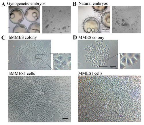
The derivation of marine medaka ESCs. (A) Marine medaka gynogenetic embryos and gynogenetic blastomeres. (B) Marine medaka normal embryos and their blastomeres. (C) hMMES cell colony and hMMES1 cells at passage 30. (D) MMES cell colony and MMES1 cells at passage 30. Scale bars, 200 μm unless otherwise indicated. All experiments were performed at least three times, and representative data are shown.

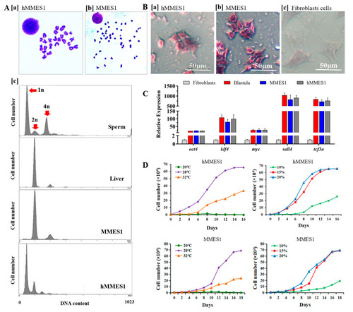
Karyotype, pluripotency, and growth curves of hMMES1. (A) Chromosome metaphase of hMMES1 [a] and MMES1 [b], respectively; [c] flow analyses of DNA content in hMMES1, MMES1, sperm, and liver cells from marine medaka. (B) Alkaline phosphatase staining of hMMES1 [a] and MMES1 [b] at day 248 of culture; the cultured fibroblasts cells [c] were stained as a control. (C) Expression of pluripotency marker genes in hMMES1, MMES1, and fibroblast cells by qRT-PCR; β-actin was used as a reference gene. Data are representative of three repeated experiments as mean ± SEM. One-way ANOVA was used for multiple-group comparisons. (D) Growth curves of hMMES1 and MMES1 cells at different temperatures in ESM4 supplemented with 15% fetal bovine serum or different concentrations of FBS at 28 °C. Representative data from three independent experiments are presented.

The formation and differentiation of hMMES1-derived embryoid bodies (EBs). (A) Morphology of 10-day-old EBs derived from hMMES1 and MMES1. (B) Expression analysis of gene markers specific to the ectoderm (nf200), mesoderm (actinin2 and ntl), and endoderm (sox17) in 10-day-old hMMES1 or MMES1-derived EBs by qRT-PCR. Data are presented as the means ± SD of three independent experiments. Student’s t-test was conducted for two-group comparisons (** p < 0.01). (C) Morphology of differentiated cells from hMMES1- or MMES1-derived EBs. (D) Expression analysis of muscle (myoD) and neural (sox10) marker genes expression in hMMES1 or MMES1 differentiated cells by qRT-PCR. Data are represented as mean ± SEM from three independent replicates. Student’s t-test was conducted for two-group comparisons (** p < 0.01). (E) Micrograph of marine medaka chimera embryo host (n = 120), showing transplanted hMMES1 (red) or MMES1 (green) at 0, 3, and 9 days post-fertilization (dpf). Scale bars, 100 μm. Representative data are shown from three independent experiments.

Interspecific chimera formation of hMMES1 in a zebrafish host. (A) Micrograph of zebrafish embryo host (n = 200) showing transplanted hMMES1 (red) and MMES1 (green) at 0, 1, and 3 dpf. Scale bars, 100 μm. (B) Molecular analyses of zebrafish transplanted with MMES1 and hMMES1, wild zebrafish, and medaka embryos at day 1–10 (D1–D10) post-fertilization. The ectoderm markers (nf200 and gfap), mesoderm markers (ntl and myf5), endoderm markers (hnf3b and sox17), and neural crest markers (mitf) were amplified by using primers specific to medaka cDNAs; β-actin of marine medaka or zebrafish was amplified as a reference. All experiments were performed three times, and representative data are shown. The uncropped nucleic acid gels figures are presented in the Supplementary Materials.

Transfection and transduction efficiency of hMMES1 cells. (A,B): hMMES1 (A) and MMES1 (B) cells were transfected with pEGFP-N3 plasmid for 48 h. Bar = 100 μm. (C,D): hMMES1 (C) and MMES1 (D) cells were infected with rvLcherry at MOI = 50 for 48 h. Bar = 100 μm. Representative images of cells are shown from three independent experiments.

Viral susceptibility of hMMES1 cells. (A) hMMES1 and MMES1 cells were infected with RGNNV or SGIV for 48 h. Bar = 100 μm. (B) Expression analysis of RGNNV or SGIV mRNA in hMMES1 and MMES1 cells at 48 h following virus infection. β-actin of marine medaka was amplified as a reference. All experiments were performed three times and shown with the representative data. (C) Transmission electron micrograph (×50,000) of RGNNV- or SGIV-infected hMMES1 or MMES1 cells; cells treated with PBS were used as a control (Three independent biological experiments, n = 3). The uncropped nucleic acid gels figures are presented in the Supplementary Materials.

Acknowledgments
This image is the copyrighted work of the attributed author or publisher, and ZFIN has permission only to display this image to its users. Additional permissions should be obtained from the applicable author or publisher of the image. Full text @ Animals (Basel)