IMAGE

Figure 3

ID
ZDB-IMAGE-240930-89
Source
Figures for Zhang et al., 2024
Image
Figure Caption

Figure 3

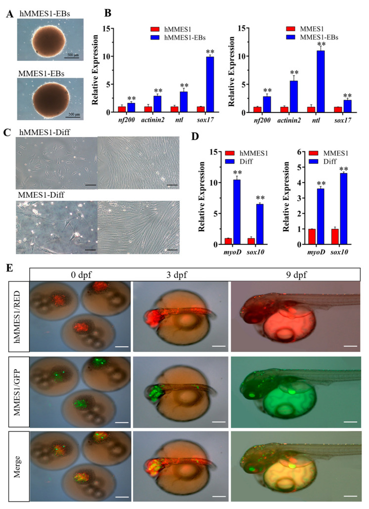
The formation and differentiation of hMMES1-derived embryoid bodies (EBs). (A) Morphology of 10-day-old EBs derived from hMMES1 and MMES1. (B) Expression analysis of gene markers specific to the ectoderm (nf200), mesoderm (actinin2 and ntl), and endoderm (sox17) in 10-day-old hMMES1 or MMES1-derived EBs by qRT-PCR. Data are presented as the means ± SD of three independent experiments. Student’s t-test was conducted for two-group comparisons (** p < 0.01). (C) Morphology of differentiated cells from hMMES1- or MMES1-derived EBs. (D) Expression analysis of muscle (myoD) and neural (sox10) marker genes expression in hMMES1 or MMES1 differentiated cells by qRT-PCR. Data are represented as mean ± SEM from three independent replicates. Student’s t-test was conducted for two-group comparisons (** p < 0.01). (E) Micrograph of marine medaka chimera embryo host (n = 120), showing transplanted hMMES1 (red) or MMES1 (green) at 0, 3, and 9 days post-fertilization (dpf). Scale bars, 100 μm. Representative data are shown from three independent experiments.

Acknowledgments
This image is the copyrighted work of the attributed author or publisher, and ZFIN has permission only to display this image to its users. Additional permissions should be obtained from the applicable author or publisher of the image. Full text @ Animals (Basel)