
- Title
-
Lemairamin (Wgx-50) Attenuates DSS-Induced Intestinal Inflammation in Zebrafish
- Authors
- Zhang, L., Liu, H., Zhang, H., Yuan, H., Ren, D.
- Source
- Full text @ Int. J. Mol. Sci.
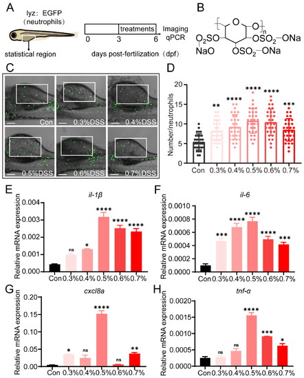
DSS promotes the recruitment of neutrophils and the expression of pro-inflammatory cytokines. (A) Tg(lyz:EGFP) zebrafish larvae (3 dpf) were exposed to different concentrations of DSS for three days. (B) Images display the chemical formula of DSS. (C) Images demonstrate the recruitment of neutrophils in the zebrafish intestinal injury model under fluorescence microscopy. White rectangles represent counting areas. Scale: 100 μm. (D) Shows the statistical quantification of neutrophil recruitment in the zebrafish intestinal injury model under control group and different concentrations of DSS treatment. The recruitment of neutrophils in the zebrafish intestine increases to varying degrees with different concentrations of DSS treatment. (E–H) Display the gene expression levels of pro-inflammatory cytokines il-1, il-6, cxcl8a, and tnf-α in zebrafish under control group and different DSS concentrations. The levels of pro-inflammatory cytokines in zebrafish increase to varying degrees with different concentrations of DSS treatment. All statistical analyses are compared to the control group (Con group). (ns, not significant; * p < 0.05, ** p < 0.01, *** p < 0.001, **** p < 0.0001 one-way ANOVA.) |
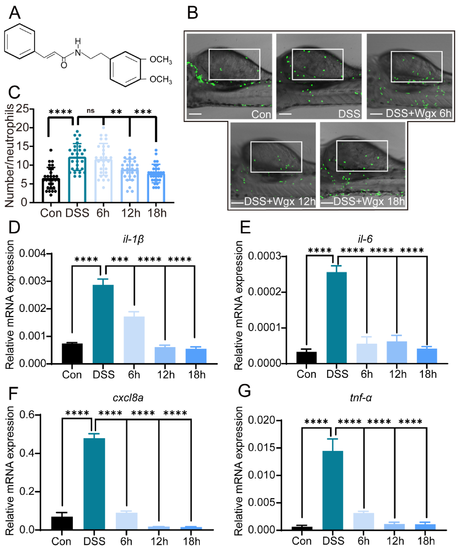
Wgx-50 inhibits the recruitment of neutrophils and the expression of pro-inflammatory cytokines induced by DSS. (A) Images display the chemical formula of Wgx-50. (B) Images demonstrate the recruitment of neutrophils in the zebrafish intestinal injury model under fluorescence microscopy. White rectangles represent counting areas. Scale: 100 μm. (C) The statistical quantification of neutrophil recruitment in zebrafish intestinal injury model under control group, DSS group, and DSS + Wgx-50 group. The recruitment of neutrophils in the zebrafish intestine increases after DSS treatment, and the number of neutrophil recruitment gradually decreases with the addition of Wgx-50 over time. (D–G) The gene expression levels of pro-inflammatory cytokines il-1, il-6, cxcl8a, and tnf-α in zebrafish under control group, DSS group, and DSS + Wgx-50 group. The levels of pro-inflammatory cytokines increase after DSS treatment, and the levels gradually decrease with the addition of Wgx-50 over time. The primer sequences are listed in Table 1. (ns, not significant;, ** p < 0.01, *** p < 0.001, **** p < 0.0001, one-way ANOVA.) |
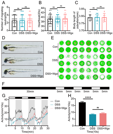
DSS and Wgx-50 have no effect on the growth and development of zebrafish. (A) The number of spontaneous movements per minute in zebrafish larvae at 30 hpe (n = 20). There was no significant effect observed after drug treatment. (B) The number of heartbeats every 20 s in zebrafish larvae at 48 hpe (n = 20). There was no significant effect observed after drug treatment. (C) The body length of zebrafish larvae at 96 hpe (n = 10/group). There was no significant effect observed after drug treatment. (D) The morphology of zebrafish at 96 hpf was captured using a stereomicroscope. Scale bar: 100 μm. (E) Zebrafish larval PMR behavior monitored by Viewpoint system (n = 16). (F) The experimental workflow of PMR is shown in the diagram. (G,H) Data analysis of zebrafish larval PMR behavior monitored by Viewpoint system (n = 16). There was a significant decrease in activity after drug treatment. (ns, not significant; **** p < 0.0001, one-way ANOVA.) |
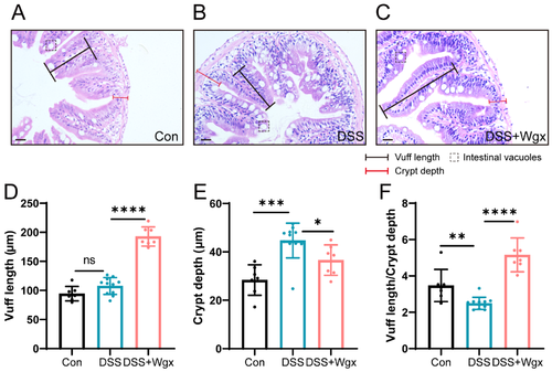
Protective effect of Wgx-50 on intestinal morphology in zebrafish after DSS treatment. (A) H&E staining of control group zebrafish intestinal sections shows normal tissue structure. (B) Zebrafish intestinal sections exposed to DSS show evident tissue inflammation and pathological changes. (C) Zebrafish intestinal sections treated with Wgx show a protective effect. Scale bar: 20 μm. (D–F) Zebrafish intestinal villus length, crypt depth, and the ratio of villus length to crypt depth in the Con, DSS, and DSS + Wgx groups. (ns, not significant; * p < 0.05, ** p < 0.01, *** p < 0.001, **** p < 0.0001, one-way ANOVA.) |
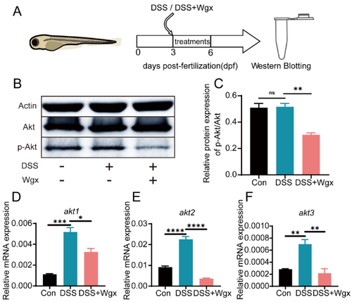
Wgx-50 affects inflammation by regulating Akt expression. (A) Zebrafish larvae (3 dpf) were exposed to DSS or DSS+Wgx on the third day and treated for three days, followed by Western blot analysis. (B,C) Protein expression levels of p-Akt/Akt in control group, DSS group, and DSS + Wgx group are shown. There was no significant change in p-Akt/Akt protein expression levels after DSS treatment, while the protein levels significantly decreased after Wgx treatment. (D–F) Gene expression levels of akt1, akt2, and akt3 in control group, DSS group, and DSS + Wgx group are shown. There was a significant upregulation of akt1, akt2, and akt3 gene expression levels after DSS treatment, while the gene levels significantly decreased after Wgx treatment. (ns, not significant; * p < 0.05, ** p < 0.01, *** p < 0.001, **** p < 0.0001, one-way ANOVA.) |