
|
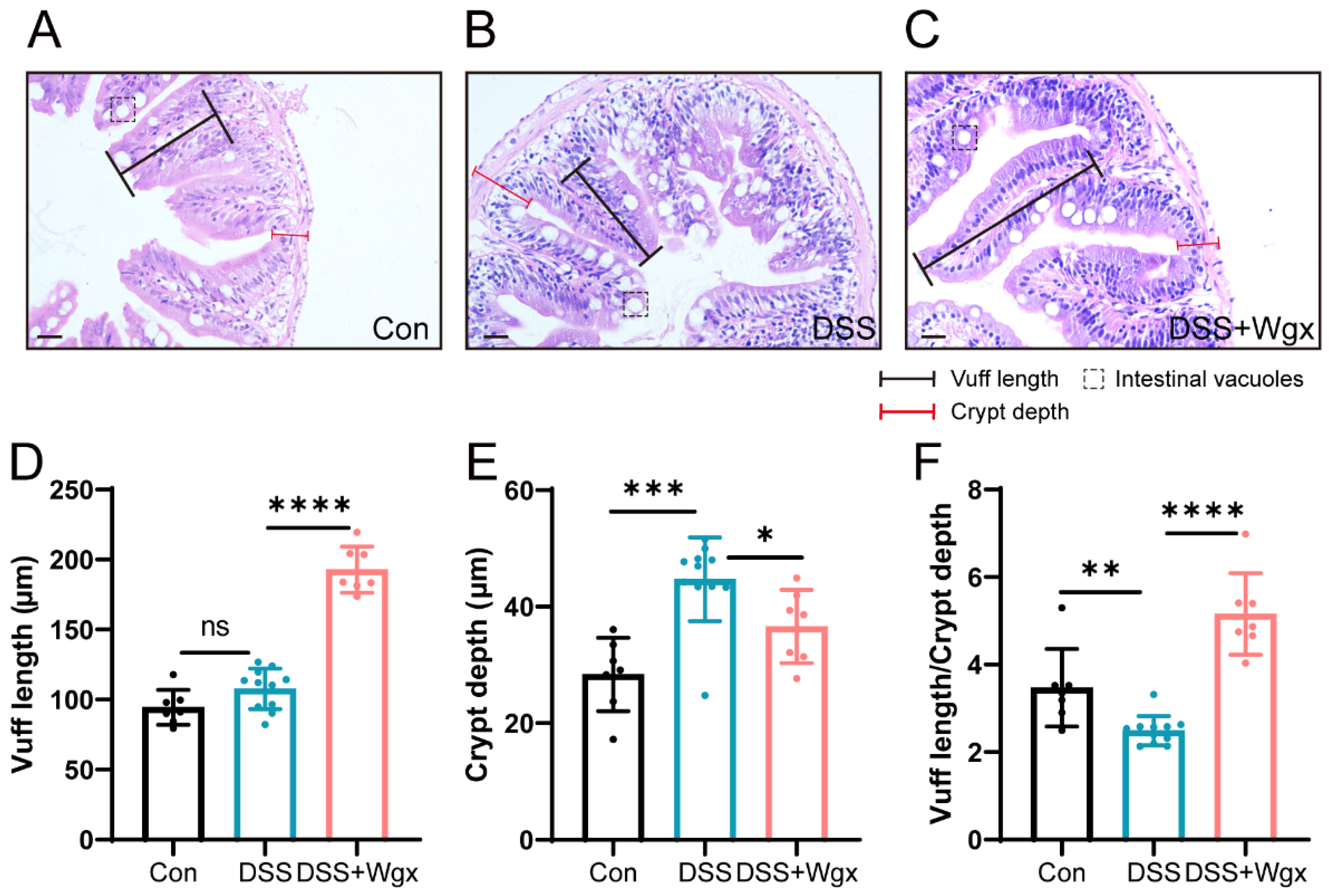
Fig. 4 Protective effect of Wgx-50 on intestinal morphology in zebrafish after DSS treatment. (A) H&E staining of control group zebrafish intestinal sections shows normal tissue structure. (B) Zebrafish intestinal sections exposed to DSS show evident tissue inflammation and pathological changes. (C) Zebrafish intestinal sections treated with Wgx show a protective effect. Scale bar: 20 μm. (D–F) Zebrafish intestinal villus length, crypt depth, and the ratio of villus length to crypt depth in the Con, DSS, and DSS + Wgx groups. (ns, not significant; * p < 0.05, ** p < 0.01, *** p < 0.001, **** p < 0.0001, one-way ANOVA.)