FIGURE SUMMARY
Title

Developmental Delay and Male-Biased Sex Ratio in esr2b Knockout Zebrafish

Authors
Peng, W., Zhang, Y., Song, B., Yang, P., Liu, L.
Source
Full text @ Genes (Basel)

Genomic structure and targeted genetic modification of esr2b in zebrafish. (A) Gene structure of esr2b. Zebrafish esr2b contains 10 exons, with the translation initiation codon located on the second exon. Deletion of 16 nucleotides results in translation frameshift and early termination. * represent translation termination. Theoretically, the esr2b mRNA of knockout zebrafish only coding 60 amino acids. (B) Verified sequence change in esr2b knockout zebrafish.

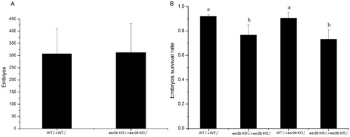
Comparison of fertility and survival rate between wild-type and esr2b knockout zebrafish. (A) The average number of embryos produced by wild-type and esr2b knockout homozygote zebrafish, (B) The survival rate of four kinds of embryos (wt♀ × wt♂, esr2b KO♀ × esr2b KO♂, wt♀ × esr2b KO♂, esr2b KO♀ × WT♂). Eight pairs from different zebrafish lines (8 male and 8 female) were selected for spawning. The results are presented as mean ± SEM values. Values accompanied by different letters are significantly different (ANOVA followed by Tukey’s test, p < 0.05).

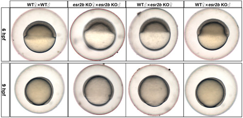
The developmental delay of
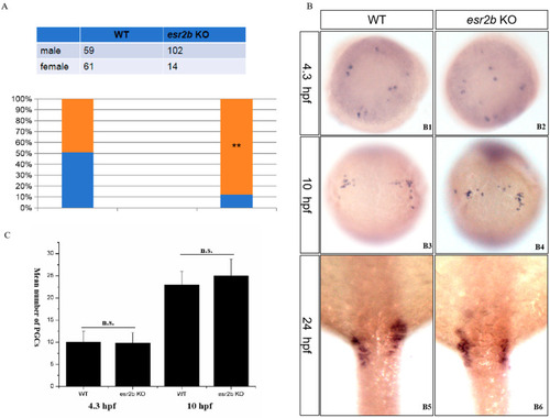
 esr2b knockout zebrafish. The developmental situation of zebrafish embryos was observed and photographed at 6 hpf and 9 hpf, respectively. All embryos from different zebrafish lines were collected and randomly transferred into Petri dishes (30/each).

The influence of esr2b knockout on sex ratio and the number of PGCs during development. (A). Sex ratios of esr2b knockout zebrafish and wild-type zebrafish at 90 dpf. Orange and blue represents the proportion of male and female zebrafish in the group, respectively. (B) Whole-mount in situ hybridization with vasa mRNA probe during early development. At 4.3 hpf, also named the dome stage, the average number of vasa signals in wild-type and esr2b KO embryos were 10 (n = 15) and 9.8 (n = 15) (B1,B2); At 10 hpf, also named the bud stage, the average number of vasa signals in wild-type and esr2b KO embryos were 22.9 (n = 15) and 25 (n = 15) (B3,B4); At 24 hpf, the vasa signal intensity showed no significant difference (B5,B6). (C) Comparison of the number of PGCs between wild-type and esr2b knockout zebrafish at 4.3 hpf and 10 hpf. n.s.; not significant. **, p < 0.01.

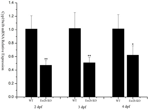
Reduced expression levels of
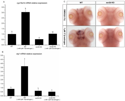
 cyp19a1b mRNA in esr2b knockout zebrafish at 2 dpf, 3 dpf and 4 dpf. (*, p < 0.05, **, p < 0.01). Group of 30 wild-type or esr2b knockout zebrafish embryos were cultured in Petri dishes (4 cm in diameter). At 2 dpf, 3 dpf and 4 dpf, four individual samples of wild-type or esr2b knockout zebrafish embryos were collected for RNA extraction. Levels of cyp19a1b mRNA relative to β-actin were detected by qPCR.

Response of cyp19a1b and vtg1 expression to estrogen stimulation. (A,B) Groups of 30 zebrafish embryos were treated from 2 to 4 dpf with 1 μM 17β-estradiol (E2). Levels of cyp19a1b and vtg1 mRNA relative to β-actin RNA were determined by qPCR. The results are presented as mean ± SEM values. Values accompanied by different letters are significantly different (ANOVA followed by Tukey’s test, p < 0.05). (C) cyp19a1b expression analysis by whole-mount in situ hybridization.

Acknowledgments
This image is the copyrighted work of the attributed author or publisher, and ZFIN has permission only to display this image to its users. Additional permissions should be obtained from the applicable author or publisher of the image. Full text @ Genes (Basel)