FIGURE SUMMARY
Title

Modulating mycobacterial envelope integrity for antibiotic synergy with benzothiazoles

Authors
Habjan, E., Lepioshkin, A., Charitou, V., Egorova, A., Kazakova, E., Ho, V.Q., Bitter, W., Makarov, V., Speer, A.
Source
Full text @ Life Sci Alliance

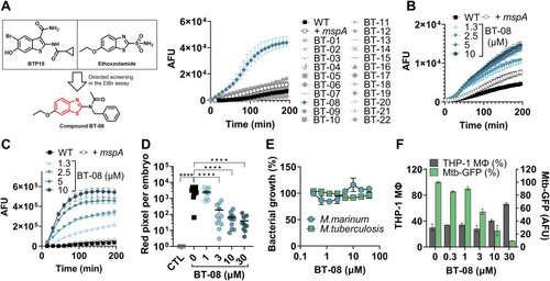
Screening for membrane permeabilizing agents identifies BT-08 as a hit compound with in vivo and ex vivo anti-mycobacterial activity.

(A) Chemical structure of BTP15 and ethoxzolamide served as a starting point to generate a benzothiazole compound library, from which BT-08 was the most promising hit. WT M. marinum cultures were grown (7H9 medium with albumin–dextrose–saline [ADS] and tyloxapol) in the presence of test compounds (10 μM) before the EtBr uptake assay was performed. A WT M. marinum strain expressing the porin mspA served as a positive control (+mspA). After the addition of EtBr, the fluorescence intensity (arbitrary fluorescence units) was measured for 3 h. Data are presented as the mean of triplicates ± range. (B) Dose-dependent activity of BT-08 on M. marinum during the ethidium bromide uptake assay (7H9 medium with ADS and tyloxapol). Data are presented as the mean of triplicates ± range. (C) Dose-dependent activity of BT-08 on M. marinum during the resazurin uptake assay (7H9 medium with ADS and tyloxapol). Data are presented as the mean of triplicates ± range. (D) Dose-dependent activity of BT-08 in the zebrafish embryo infection model. Embryos were yolk-infected with M. marinum expressing tdTomato, and treatment was performed by immersion. Each data point represents the integrated red fluorescence intensity of a single zebrafish embryo, and the signal of each group is expressed as the mean ± SD of the mean. Statistical significance was determined by one-way ANOVA, following Dunnett’s multiple comparison test by comparing the signal from the DMSO-treated control sample with each treatment group (****P ≤ 0.0001). CTL represents the non-infected group. (E) Effect of BT-08 on bacterial growth of M. marinum MUSA and M. tuberculosis H37Rv was assessed using the resazurin reduction microtiter plate assay. DMSO-treated sample represents 100% bacterial growth. Data are presented as the mean of duplicates ± range. (F) Intracellular activity of BT-08 in THP-1 macrophages infected with M. tuberculosis expressing gfp. The expression of gfp was induced by the addition of ATc. To detect macrophages (gray bars), the nuclei were stained with Hoechst dye. The GFP signal within each macrophage was quantified, representing the amount of viable bacteria (green bars). DMSO- and rifampicin (10 μM)-treated samples served as a negative and positive control, respectively. Data are presented as the mean of duplicates ± range.

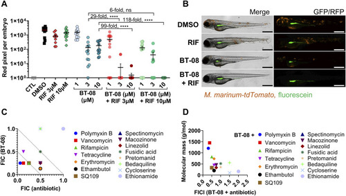
Compound BT-08 and high molecular weight antibiotics act synergistically in vivo and in vitro.

(A) Activity of rifampicin (RIF) and BT-08 and their combinations in the zebrafish embryo infection model. Zebrafish embryos were yolk-infected with M. marinum expressing tdTomato, and treatment was performed by immersion. Each data point represents the integrated red fluorescence intensity of a single zebrafish embryo, and the signal of each group is expressed as the mean ± SD of the mean. CTL represents the non-infected group. The fold difference between the means of different treatment groups is depicted. Statistical significance was determined by one-way ANOVA, following Tukey’s multiple comparison test by comparing each treatment group with the rest (****P ≤ 0.0001). CTL represents the non-infected group. (B) Representative images of M. marinum yolk-infected zebrafish embryos treated with DMSO, BT-08 at 3 μM, RIF at 3 μM, or their combination (BT-08 at 3 μM and RIF at 3 μM) at 4 d post-fertilization. The red color corresponds to a signal from M. marinum expressing tdTomato, and the green color corresponds to the injection control dye fluorescein. The scale bar represents 500 μm. (C) Drug combinations between BT-08 and selected antibiotics reported as the fractional inhibitory concentration (FIC) and the FIC index (FICI) were investigated in M. marinum using a checkerboard assay using the HdB medium with Tween-80. (D) FICI of BT-08 and selected antibiotics calculated using a checkerboard assay in M. marinum (HdB medium with Tween-80) is plotted against the molecular weight (g/mol) of tested antibiotics. The correlation was evaluated using Spearman’s one-tailed correlation test, showing significance of P = 0.0315.

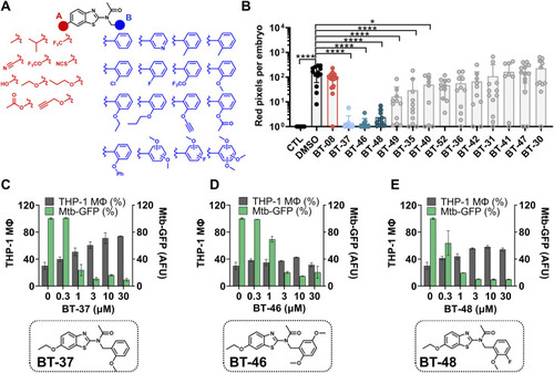
Structure–activity relationship studies revealed the most active derivative BT-37.

(A) Set of benzothiazole derivatives for structure–activity relationship studies. (B) Activity of benzothiazole derivatives at 1 μM in the zebrafish–M. marinum infection model. Embryos were yolk-infected with M. marinum expressing tdTomato and treated with 1 μM of each derivative by immersion. Each data point represents the integrated red fluorescence intensity of a single zebrafish embryo, and the signal of each group is expressed as the mean ± SD of the mean. Statistical significance was determined by one-way ANOVA, following Dunnett’s multiple comparison test by comparing the signal from the DMSO-treated control sample with each treatment group (****P ≤ 0.0001; *P ≤ 0.05). CTL represents the non-infected group. (C, D, E) Activity and chemical structures of BT-37, (D) BT-46, and (E) BT-48 in the macrophage infection model. THP-1 macrophages were infected with M. tuberculosis expressing gfp, induced by adding ATc. Macrophages (gray bars) were detected by staining the nuclei with Hoechst dye. The GFP signal within each macrophage was quantified, representing the amount of viable bacteria (green bars). DMSO- and RIF (10 μM)-treated samples served as a negative and positive control, respectively. Data are presented as the mean of duplicates ± range.

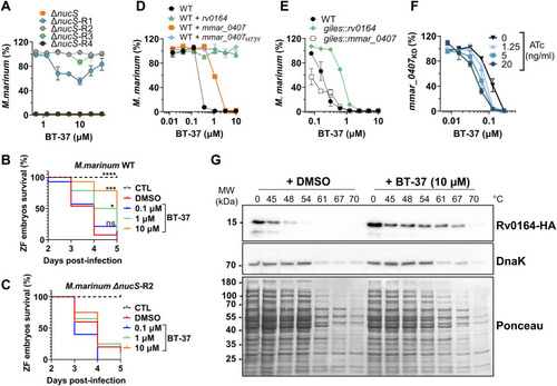
Mutations and expression level of MMAR_0407 (Rv0164) modulate susceptibility to BT-37.

(A) Spontaneous BT-37–resistant mutants of hyper-mutating M. marinum ΔnucS strain were tested for their susceptibility toward BT-37 using the resazurin microtiter plate assay in the HdB medium with Tween-80. Data are presented as the mean of duplicates ± range. (B, C) Survival of zebrafish embryos that were yolk-infected with the M. marinum WT or (C) BT-37–resistant M. marinum strain (M. marinum ΔnucS-R2) after dose-dependent treatment with BT-37. Kaplan–Meier’s survival tests were conducted to generate the survival curves, and P-values were calculated by the log-rank test (*P = 0.0173; ***P = 0.0001; ****P ˂ 0.0001). CTL: non-infected control sample. (D) Susceptibility of the M. marinum WT strain or strains transformed with episomal plasmids pMN016-rv0164 (WT+rv0164), pMN016-mmar_0407 (WT+mmar_0407), or pMN016-mmar_0407H73Y (WT+mmar_0407H73Y) toward compound BT-37 after 4 d of incubation in the HdB medium with Tween-80 using the resazurin microtiter plate assay. Data are presented as the mean of duplicates ± range. (E) Susceptibility of the M. marinum WT strain or strains transformed with an integrative plasmid pML1357-mmar_0407 (giles::mmar_0407) or pML1357-rv0164 (giles::rv0164) toward BT-37 in the HdB medium with Tween-80 using the resazurin microtiter plate assay. Data are presented as the mean of duplicates ± range. (F)M. marinum strain expressing mmar_0407-targeting sgRNA (M. marinum mmar_0407KD) was incubated with a range of concentrations of ATc and BT-37 in the HdB medium with Tween-80. The susceptibility of this ATc-inducible knock-down strain was measured using the resazurin microtiter plate assay. Data are presented as the mean of duplicates ± range. (G) Thermal shift assay of Rv0164-HA, which was expressed in E. coli. Cell lysates were incubated with BT-37 or DMSO and exposed to different temperatures. Denatured proteins were removed by centrifugation, and the remaining proteins were separated by SDS–PAGE (Ponceau) and analyzed by Western blotting. DnaK served as an internal control.

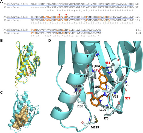
Molecular docking simulation of the structure of the Rv0164 complex with BT-37.

(A) Protein sequence alignment of MMAR_0407 in M. marinum and Rv0164 in M. tuberculosis using Clustal Omega. An asterisk (*) indicates positions that have identical residues, a colon (:) indicates that the residues have conserved properties, and a dot (.) indicates residues that are semi-conserved. Amino acids highlighted in orange are in 2 Å vicinity of the compound BT-37 and contribute to inhibitor binding. Red asterisks indicate residues contributing to experimentally confirmed resistance in M. marinum (Mmar G69/Mtb G77 and Mmar H73/Mtb H81). (B) Structure alignment of MMAR_0407 in M. marinum (yellow) and Rv0164 in M. tuberculosis (cyan). The RMSD score between these structures is 0.158 Å. In orange is depicted the compound BT-37 as the top model of docking simulation, according to the HADDOCK score. (C) Definition of the top-scored binding pocket in Rv0164 that was used to define the binding information of the docking simulation. (D) Top model of the docking simulation, according to the HADDOCK score, is shown in more detail. The corresponding residues in M. tuberculosis (G77, H81) contributing to experimentally confirmed resistance in M. marinum are labeled in red.

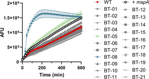
Compound BT-08 increases resazurin uptake.

WT M. marinum cultures were grown (7H9 medium with albumin–dextrose–saline and tyloxapol) in the presence of test compounds (10 μM) before the resazurin uptake assay was performed. A WT M. marinum strain expressing the porin mspA served as a positive control (+mspA). After the addition of resazurin sodium salt mixed with Tween-80, the fluorescence intensity (arbitrary fluorescence units) was measured for 10 h. Data are presented as the mean of triplicates ± SD.

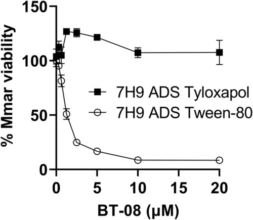
Detergents used in growth media affect the susceptibility of M. marinum to BT-08.

Susceptibility of M. marinum toward BT-08 in 7H9 medium supplemented with albumin–dextrose–saline and different detergents: Tween-80 or tyloxapol, using the resazurin microtiter plate assay. Data are presented as the mean of duplicates ± SD.

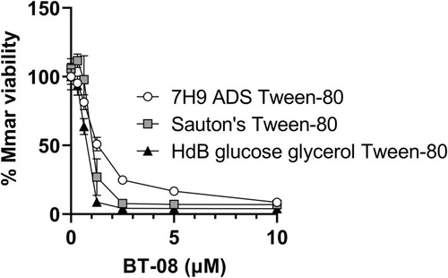
Different growth media affect susceptibility of M. marinum to BT-08.

Susceptibility of M. marinum toward BT-08 in different growth media using the resazurin microtiter plate assay. Data are presented as the mean of duplicates ± SD.

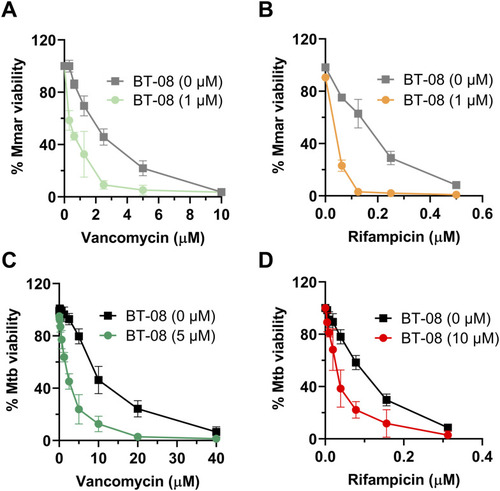
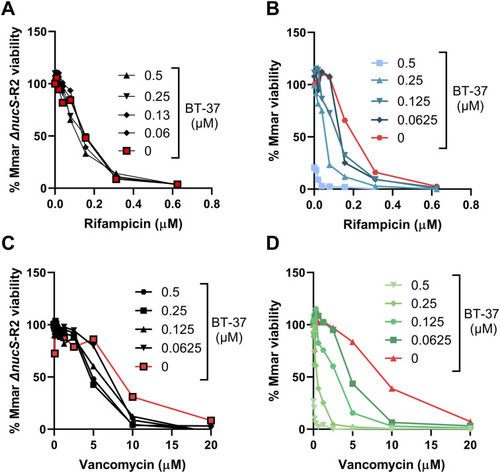
Compound BT-08 increases activity of rifampicin (RIF) and vancomycin in vitro.

(A, B, C, D) Sensitivity of M. marinum toward the combinatorial treatment of compound BT-08 with vancomycin (A) or RIF, (B, C, D) and the sensitivity of M. tuberculosis toward the combinatorial treatment of compound BT-08 with vancomycin (C) or RIF (D) using the in vitro checkerboard assay and 7H9 media supplemented with albumin–dextrose–saline and tyloxapol. Data are presented as the mean of duplicates ± SD.

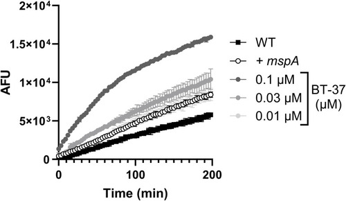
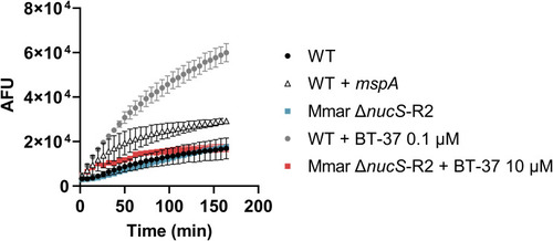
Treatment of M. marinum with BT-37 increases ethidium bromide (EtBr) uptake.

The M. marinum strain was grown in the HdB medium with Tween-80 and with different concentrations of BT-37. M. marinum WT expressing the porin mspA was used as a positive control (+mspA). After adding EtBr, the fluorescence intensity (arbitrary fluorescence units) was measured for 3 h. Data are presented as the mean of triplicates ± SD.

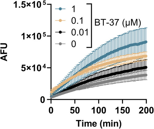
Treatment of M. tuberculosis with BT-37 increases ethidium bromide (EtBr) uptake.

The
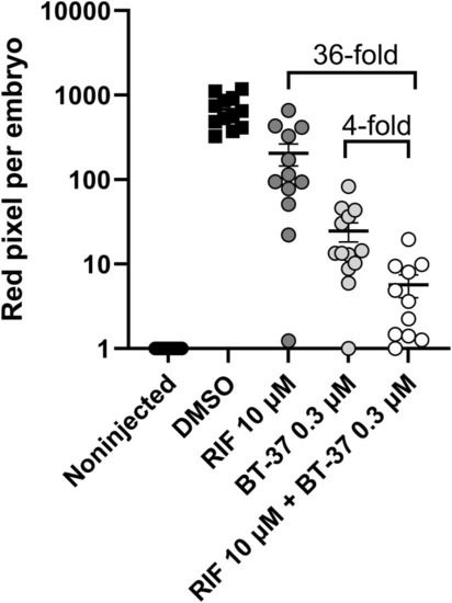
 M. tuberculosis mc26206 strain was grown in the HdB medium supplemented with pantothenic acid, leucine, and Tween-80, and with different concentrations of BT-37. After adding EtBr, the fluorescence intensity (arbitrary fluorescence units) was measured for 3 h. Data are presented as the mean of duplicates ± SD.

Synergistic effect of RIF and BT-37 in M. marinum-infected zebrafish embryos

The activity of rifampicin at 10 μM and BT-37 at 0.3 μM and their combination in the zebrafish embryo infection model. Zebrafish embryos were yolk-infected with M. marinum expressing tdTomato, and treatment was performed by immersion. Each data point represents the integrated red fluorescence intensity of a single zebrafish embryo, and the signal of each group is expressed as the mean ± SD of the mean. The fold difference between the means of different treatment groups is depicted.

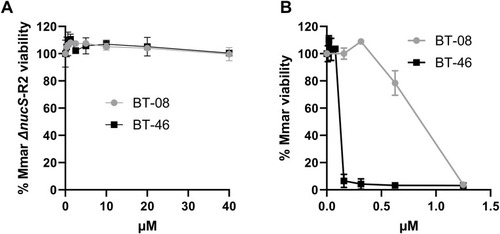
BT-08 and BT-46 exhibit cross-resistance to the BT-37–resistant M. marinum strain.

(A, B) Susceptibility of M. marinum BT-37–resistant isolates (Mmar ΔnucS-R2) and (B) M. marinum WT strain using the resazurin microtiter plate assay and the HdB medium with Tween-80. Data are presented as the mean of duplicates ± SD.

BT-37–resistant M. marinum strain does not show synergy with vancomycin or rifampicin in vitro

(A, B, C, D)M. marinum WT strain or BT-37–resistant strain (Mmar ΔnucS-R2) was investigated for the sensitivity toward the combinatorial treatment of compound BT-37 with rifampicin (A, B) or vancomycin (C, D) using the in vitro checkerboard assay and the HdB medium with Tween-80.

Treatment of BT-37–resistant M. marinum does not increases ethidium bromide (EtBr) uptake.

The M. marinum WT strain or BT-37–resistant M. marinum (Mmar ΔnucS-R2) were grown in the HdB medium with Tween-80 and with different concentrations of BT-37. After adding EtBr, the fluorescence intensity (arbitrary fluorescence units) was measured for 3 h. Data are presented as the mean of duplicates ± SD.

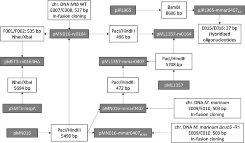
Cloning strategy for new plasmids used in this study.

Constructed plasmids are included in gray boxes. The primer pairs used for PCR amplification and correlated restriction enzymes for cloning are listed in white boxes. The DNA template for the PCRs is listed above the primer pairs. If several primer pairs are listed, overlap PCR was used to fuse the PCR fragments. When a DNA fragment was obtained by digestion of a plasmid, the used restriction enzymes and the length of the obtained fragments are indicated. Constructed plasmids with their features and primers with their sequences are listed in Tables S9 and S10, respectively.

Acknowledgments
This image is the copyrighted work of the attributed author or publisher, and ZFIN has permission only to display this image to its users. Additional permissions should be obtained from the applicable author or publisher of the image. Full text @ Life Sci Alliance