IMAGE

Figure 3.

ID
ZDB-IMAGE-240518-8
Source
Figures for Habjan et al., 2024
Image
Figure Caption

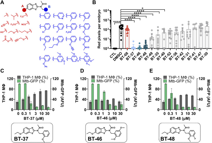
Figure 3. Structure–activity relationship studies revealed the most active derivative BT-37.

(A) Set of benzothiazole derivatives for structure–activity relationship studies. (B) Activity of benzothiazole derivatives at 1 μM in the zebrafish–M. marinum infection model. Embryos were yolk-infected with M. marinum expressing tdTomato and treated with 1 μM of each derivative by immersion. Each data point represents the integrated red fluorescence intensity of a single zebrafish embryo, and the signal of each group is expressed as the mean ± SD of the mean. Statistical significance was determined by one-way ANOVA, following Dunnett’s multiple comparison test by comparing the signal from the DMSO-treated control sample with each treatment group (****P ≤ 0.0001; *P ≤ 0.05). CTL represents the non-infected group. (C, D, E) Activity and chemical structures of BT-37, (D) BT-46, and (E) BT-48 in the macrophage infection model. THP-1 macrophages were infected with M. tuberculosis expressing gfp, induced by adding ATc. Macrophages (gray bars) were detected by staining the nuclei with Hoechst dye. The GFP signal within each macrophage was quantified, representing the amount of viable bacteria (green bars). DMSO- and RIF (10 μM)-treated samples served as a negative and positive control, respectively. Data are presented as the mean of duplicates ± range.

Acknowledgments
This image is the copyrighted work of the attributed author or publisher, and ZFIN has permission only to display this image to its users. Additional permissions should be obtained from the applicable author or publisher of the image. Full text @ Life Sci Alliance