IMAGE

Figure 4.

ID
ZDB-IMAGE-240518-12
Source
Figures for Habjan et al., 2024
Image
Figure Caption

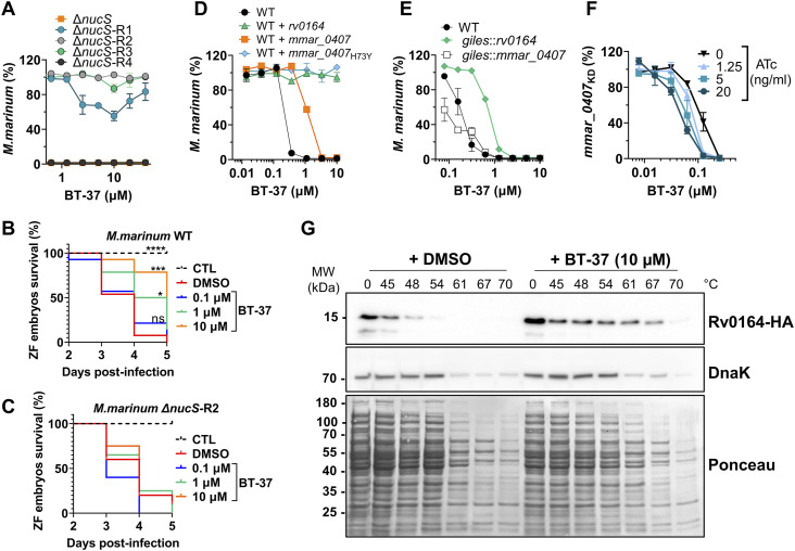
Figure 4. Mutations and expression level of MMAR_0407 (Rv0164) modulate susceptibility to BT-37.

(A) Spontaneous BT-37–resistant mutants of hyper-mutating M. marinum ΔnucS strain were tested for their susceptibility toward BT-37 using the resazurin microtiter plate assay in the HdB medium with Tween-80. Data are presented as the mean of duplicates ± range. (B, C) Survival of zebrafish embryos that were yolk-infected with the M. marinum WT or (C) BT-37–resistant M. marinum strain (M. marinum ΔnucS-R2) after dose-dependent treatment with BT-37. Kaplan–Meier’s survival tests were conducted to generate the survival curves, and P-values were calculated by the log-rank test (*P = 0.0173; ***P = 0.0001; ****P ˂ 0.0001). CTL: non-infected control sample. (D) Susceptibility of the M. marinum WT strain or strains transformed with episomal plasmids pMN016-rv0164 (WT+rv0164), pMN016-mmar_0407 (WT+mmar_0407), or pMN016-mmar_0407H73Y (WT+mmar_0407H73Y) toward compound BT-37 after 4 d of incubation in the HdB medium with Tween-80 using the resazurin microtiter plate assay. Data are presented as the mean of duplicates ± range. (E) Susceptibility of the M. marinum WT strain or strains transformed with an integrative plasmid pML1357-mmar_0407 (giles::mmar_0407) or pML1357-rv0164 (giles::rv0164) toward BT-37 in the HdB medium with Tween-80 using the resazurin microtiter plate assay. Data are presented as the mean of duplicates ± range. (F)M. marinum strain expressing mmar_0407-targeting sgRNA (M. marinum mmar_0407KD) was incubated with a range of concentrations of ATc and BT-37 in the HdB medium with Tween-80. The susceptibility of this ATc-inducible knock-down strain was measured using the resazurin microtiter plate assay. Data are presented as the mean of duplicates ± range. (G) Thermal shift assay of Rv0164-HA, which was expressed in E. coli. Cell lysates were incubated with BT-37 or DMSO and exposed to different temperatures. Denatured proteins were removed by centrifugation, and the remaining proteins were separated by SDS–PAGE (Ponceau) and analyzed by Western blotting. DnaK served as an internal control.

Acknowledgments
This image is the copyrighted work of the attributed author or publisher, and ZFIN has permission only to display this image to its users. Additional permissions should be obtained from the applicable author or publisher of the image. Full text @ Life Sci Alliance